. 2017 Mar 21;8(12):20042-20055.
doi: 10.18632/oncotarget.15373.

Thymol mitigates lipopolysaccharide-induced endometritis by regulating the TLR4- and ROS-mediated NF-κB signaling pathways

Affiliations

Thymol mitigates lipopolysaccharide-induced endometritis by regulating the TLR4- and ROS-mediated NF-κB signaling pathways

Haichong Wu et al. Oncotarget. .

Abstract

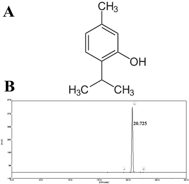
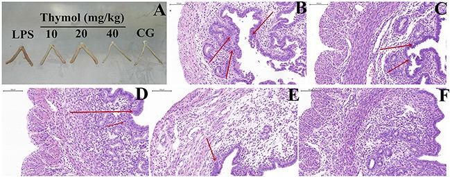
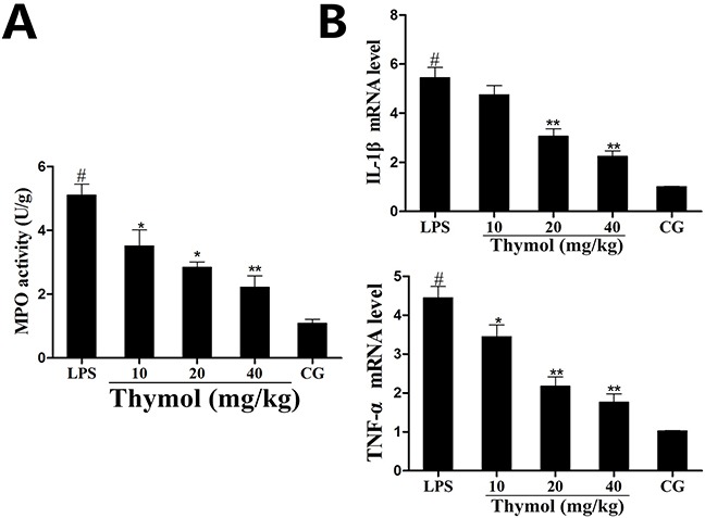
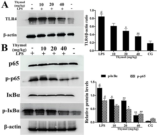
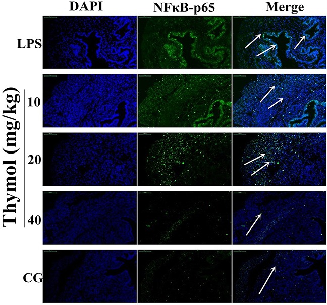
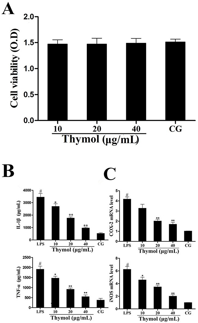
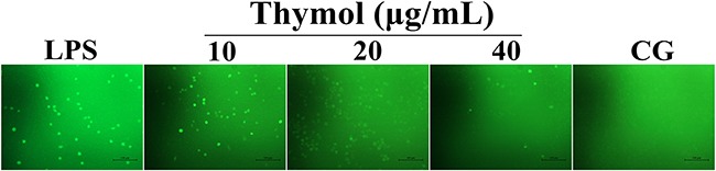
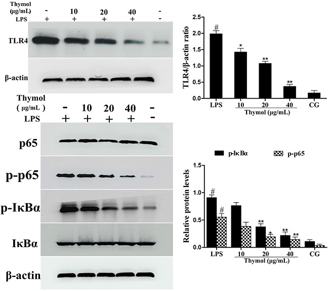
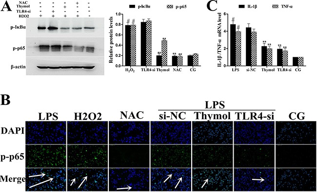
The purpose of this study was to investigate the effects of thymol on lipopolysaccharide (LPS)-induced inflammatory responses and to clarify the potential mechanism of these effects. LPS-induced mouse endometritis was used to confirm the anti-inflammatory action of thymol in vivo. RAW264.7 cells were used to examine the molecular mechanism and targets of thymol in vitro. In vivo, thymol markedly alleviated LPS-induced pathological injury, myeloperoxidase (MPO) activity, and the production of tumor necrosis factor-α (TNF-α) and interleukin-1β (IL-1β) in mice. Further studies were performed to examine the expression of the Toll-like receptor 4 (TLR4) -mediated nuclear factor-κB (NF-κB) pathway. These results showed that the expression of the TLR4-mediated NF-κB pathway was inhibited by thymol treatment. In vitro, we observed that thymol dose-dependently inhibited the expression of TNF-α, IL-1β, inducible nitric oxide synthase (iNOS), and cyclooxygenase-2 (COX-2) in LPS-stimulated RAW264.7 cells. Moreover, the results obtained from immunofluorescence assays also indicated that thymol dose-dependently suppressed LPS-induced reactive oxygen species (ROS) production. Silencing of TLR4 inhibited NF-κB pathway activation. Furthermore, H2O2 treatment increased the phosphorylation of p65 and IκBα, which were decreased when treated with N-acetyl cysteine or thymol. In conclusion, the anti-inflammatory effects of thymol are associated with activation of the TLR4 or ROS signaling pathways, contributing to NF-κB activation, thereby alleviating LPS-induced oxidative and inflammatory responses.

Keywords: TLR4; inflammation; nuclear factor-κB; reactive oxygen species; thymol.


Conflict of interest statement

CONFLICTS OF INTERESTS

The authors declare that they have no conflicts of interest.

Figures

Figure 1
Figure 1. A. Chemical structure of thymol, B. HPLC chromatogram of thymol
Figure 2
Figure 2. Effects of thymol on LPS-stimulated uterine injury
A. Morphology of the uterus. B. LPS group, C, D, E. LPS + Thymol (10, 20, and 40 mg/kg, respectively) treatment groups, F. Control group. CG indicates the control group. LPS indicates the LPS-stimulated group. The red arrow indicates the tissue lesion area. Data represent the mean ± S.E.M. of three independent experiments. #p<0.01 vs. Control group. *p<0.05 vs. LPS group. **p<0.01 vs. LPS group.
Figure 3
Figure 3. Effects of thymol on MPO activity and cytokine expression
A. MPO activity. B. Expression of TNF-α and IL-1β mRNA in tissues. GAPDH serves as the control. CG indicates the control group. LPS indicates the LPS-stimulated group. Data represent the mean ± S.E.M. of three independent experiments. #p<0.01 vs. Control group. *p<0.05 vs. LPS group, **p<0.01 vs. LPS group.
Figure 4
Figure 4. Effects of thymol on TLR4 expression and NF-κB pathway activation
A. TLR4 protein expression levels in uterine tissues. B. Expression of the p65 and IκBα proteins in uterine tissues. Phosphorylation of p65 and IκBα was analyzed using phospho-specific antibodies. β-actin served as an internal control. CG indicates the control group. LPS indicates the LPS-stimulated group. Data represent the mean ± S.E.M. of three independent experiments. #p<0.01 vs. Control group. *p<0.05 vs. LPS group, **p<0.01 vs. LPS group.
Figure 5
Figure 5. Effects of thymol on NF-κB p65 translocation into the nucleus
Paraffin-embedded uterine tissue sections were used to detect p65 translocation to the nucleus using immunofluorescence. A. Control group, B. LPS group, C, D, E. LPS + Thymol (10, 20, and 40 mg/kg, respectively) treatment groups. The white arrow indicates the translocation of p65. Data represent the mean ± S.E.M. of three independent experiments. #p<0.01 vs. Control group. *p<0.05 vs. LPS group, **p<0.01 vs. LPS group.
Figure 6
Figure 6. A. Effect of thymol on the cell viability of RAW264.7 cells
B. Effects of thymol on the expression of TNF-α and IL-1β in LPS-stimulated RAW264.7 cells. C. Expression of iNOS and COX-2 mRNA in LPS-stimulated RAW264.7 cells. GAPDH served as the control. CG indicates the control group. LPS indicates the LPS-stimulated group. Data represent the mean ± S.E.M. of three independent experiments. #p<0.01 vs. Control group. *p<0.05 vs. LPS group, **p<0.01 vs. LPS group.
Figure 7
Figure 7. Effect of thymol on ROS production in LPS-stimulated RAW264.7 cells
Microphotographs of ROS levels in LPS-stimulated RAW264.7 cells were obtained using fluorescence microscopy after DCF-DA staining (400× magnification, scale bar = 50 μm).
Figure 8
Figure 8. Effects of thymol on TLR4-mediated NF-κB pathway activation
A. Expression of TLR4 in LPS-stimulated RAW264.7 cells. B. Expression of IκBα and p65 protein in LPS-stimulated RAW264.7 cells. Phosphorylation of IκBα and p65 were analyzed using phospho-specific antibodies. β-actin served as an internal control. CG indicates the control group. LPS indicates the LPS-stimulated group. Data represent the mean ± S.E.M. of three independent experiments. #p<0.01 vs. Control group. *p<0.05 vs. LPS group, **p<0.01 vs. LPS group.
Figure 9
Figure 9. Analysis of TLR4-mediated NF-κB pathway expression in LPS-stimulated RAW264.7 cells
A. The interfering efficiency of TLR4 siRNA was estimated using qPCR. GAPDH served as a control. B. Phosphorylation levels of p65 and IκBα were examined by western blotting after silencing of TLR4 using siRNA or thymol treatment in LPS-stimulated RAW264.7 cells. CG indicates the control group. LPS indicates the LPS-stimulated group. TLR4-si indicates the TLR4 siRNA group. si-NC indicates the siRNA-negative control group. Data represent the mean ± S.E.M. of three independent experiments. #p<0.01 vs. Control group. *p<0.05 vs. LPS group, **p<0.01 vs. LPS group.
Figure 10
Figure 10. A. Effects of H2O2 on NF-κB activation after silencing of TLR4 in RAW264.7 cells
Phosphorylation levels of p65 and IκBα were determined by western blotting after knockdown of TLR4 using siRNA or thymol, NAC pre-treatment. β-actin serves as an internal control. B. Translocation of NF-κB p65 in TLR4-si RAW264.7 cells challenged with LPS as assessed using immunofluorescence. C. Expression of the cytokines, IL-1β and TNF-α, was detected using qPCR. GAPDH served as a control. CG indicates the control group. LPS indicates the LPS-stimulated group. TLR4-si indicates the TLR4 siRNA group. NAC indicates the N-acetyl cysteine group. The white arrow indicates the translocation of p65. Data represent the mean ± S.E.M. of three independent experiments. #p<0.01 vs. Control group. *p<0.05 vs. LPS group, **p<0.01 vs. LPS group.

References

    1. Eslami M, Bolourchi M, Seifi HA, Asadi F, Akbari R. Treatment of clinical endometritis in dairy cows by previously used controlled internal drug release devices. Theriogenology. 2015;84:437–445. - PubMed
    1. Sheldon IM, Price SB, Cronin J, Gilbert RO, Gadsby JE. Mechanisms of infertility associated with clinical and subclinical endometritis in high producing dairy cattle. Reproduction in domestic animals. 2009;44(Suppl 3):1–9. - PubMed
    1. Luo J, Xu Y, Zhang M, Gao L, Fang C, Zhou C. Magnolol inhibits LPS-induced inflammatory response in uterine epithelial cells : magnolol inhibits LPS-induced inflammatory response. Inflammation. 2013;36:997–1003. - PubMed
    1. Swangchan-Uthai T, Lavender CR, Cheng Z, Fouladi-Nashta AA, Wathes DC. Time course of defense mechanisms in bovine endometrium in response to lipopolysaccharide. Biology of reproduction. 2012;87:135. - PubMed
    1. Xiang NL, Liu J, Liao YJ, Huang YW, Wu Z, Bai ZQ, Lin X, Zhang JH. Abrogating ClC-3 Inhibits LPS-induced Inflammation via Blocking the TLR4/NF-kappaB Pathway. Scientific reports. 2016;6:27583. - PMC - PubMed

MeSH terms