. 2017 Mar 21;8(12):20103-20112.
doi: 10.18632/oncotarget.15384.

Ring finger protein 6 promotes breast cancer cell proliferation by stabilizing estrogen receptor alpha

Affiliations

Ring finger protein 6 promotes breast cancer cell proliferation by stabilizing estrogen receptor alpha

Yuanying Zeng et al. Oncotarget. .

Abstract

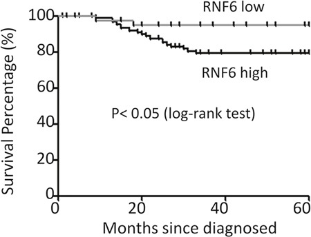
Ring finger protein 6 (RNF6) is a key oncogene in both prostate cancer and leukemia, but its role is elusive in breast cancer. In the present study, we found that RNF6 was overexpressed in more than 70% of breast cancer tissues and it was associated with overall survival. RNF6 increased breast cancer cell proliferation, migration and reduced cell sensitivity to doxorubicin. Further studies showed that RNF6 was closely associated with increased expression of estrogen receptor, a critical factor in the development of breast cancers. RNF6 was found to induce ERα expression and increased its stability. In doxorubicin-resistant breast cancer cells, RNF6 was found to be elevated in association with increased ERα and anti-apoptotic Bcl-xL, but not pro-apoptotic Bim-1. In consistence with this finding, overexpression of ERα led to increased Bcl-xL but had no effects on Bim-1. Therefore, this study demonstrated that there exists an RNF6/ERα/Bcl-xL axle in breast cancer which promotes cancer cell proliferation and survival. Targeting the RNF6/ERα/Bcl-xL axle could be a promising strategy in the treatment of breast cancer.

Keywords: Bcl-xL; ERα; breast cancer; doxorubicin; ring finger protein 6.


Conflict of interest statement

CONFLICTS OF INTEREST

The authors declare that they have no competing interest.

Figures

Figure 1
Figure 1. RNF6 is overexpressed in breast cancer
A. Fresh primary breast cancer tissues and individual para-cancerous tissues were analyzed for RNF6 expression by qRT-PCR. B. RNF6 was detected by RT-PCR using tissues from representative patients as listed in A. C: Cancer tissues; P: para-cancerous tissues. C. Statistical analysis of human breast cancer tissue arrays (n=136) stained with an anti-RNF6 antibody. Immunostaining scores (mean ± SD) for RNF6 in para-cancerous (P) and cancerous (C) tissues were summarized. D. Representative fields of human breast cancer tissue arrays by immunohistochemical staining using an anti-RNF6 antibody. E. The whole-cell lysates from a normal breast cell line and various breast cancer cell lines were extracted, followed by detecting RNF6 protein levels measured by immunoblotting analyses. GAPDH was used as a loading control.*p<0.05, ** p<0.01.
Figure 2
Figure 2. RNF6 is a negative index for the survival of breast cancer patients
The survival periods of breast cancer patients were estimated using the Kaplan-Meier estimates as described previously (see Methods). All patients were classified into two groups based on the RNF6 expression level. Estimated survival percentage of each group of patients was calculated.
Figure 3
Figure 3. RNF6 promotes breast cancer cell proliferation in MCF-7 cells
A. MCF-7 cells were transfected with a RNF6 plasmid or empty vector. Cells were then plated into 96-well plates and continued to culture for 24, 48 or 72 hrs before cell number counting. B. MCF-7 cells were infected with lentiviral shRNA of RNF6 (shRNF6) or negative control (shNC). Cell viability was evaluated at Day 1,3, and 5, respectively. C and D. RNF6-transfected or control MCF-7 cells were scratched with a tip, followed by culture in a 37°C incubator for 24 or 48 hrs. The wound gaps were analyzed by a microscope (C) followed by statistical analysis (D).
Figure 4
Figure 4. RNF6 increases MCF-7 resistance to anti-cancer agents
A. MCF-7 cells were treated with increasing concentrations of doxorubicin (ADR) or A5HQ for 24 hrs, followed by cell lysate preparation and immunoblotting against RNF6 or GAPDH. B and C. MCF-7 was treated with ADR (16 M) or 5AHQ (20 μM) for indicated time points, followed by cell lysate preparation and immunoblotting against RNF6 or GAPDH. D and E. MCF-7 cells transfected with RNF6 plasmid or empty vector (as shown in Figure 1A) for 24 hrs, followed by treatment with 5AHQ (D) or ADR (E) at indicated concentrations for 48 hrs. Cell viability was measured by MTT.
Figure 5
Figure 5. RNF6 increases the protein level of ERα
A. MCF-7 cells were treated with increased ADR or 5AHQ for 24 hrs, followed by cell lysate preparation and immunoblotting assay with specific antibodies as indicated. These blots were stripped from Figure 4A and then subjected to analysis for ERα and GAPDH. B. RNF6 and ERα were co-tranfected into HEK293 cells. Forty-eight hours later, cells were lysed for immunoblotting analysis. C. MCF-7 cells were transfected with increased RNF6 plasmids, followed by immunoblotting against ERα, Flag and GAPDH. D. MCF-7 cells were transfected with Flag-RNF6 or vector for 24 hrs, followed by CHX chase assay. Immunoblotting analysis was performed against ERα and Flag. GAPDH was used as a loading control. E. Statistically analysis of Figure D.
Figure 6
Figure 6. RNF6 promotes the ERα/Bcl-xL axle in breast cancer
A. ADR-resistant MCF-7 (MCF-7R) cells were measured associated gene expression. B. MCF-7 cells were transfected with increased Myc-ERα plasmids for 48 hrs, followed by immunoblotting analysis. C. Wild-type or RING-deficient RNF6 plasmids were co-transfected with Myc-ERα in HEK293 cells. Forty-eight hours later, cells were harvest for immunoblotting analysis against Myc and Flag. GAPDH was used as a loading control.

References

    1. McGuire S. Adv Nutr. Vol. 7. Geneva, Switzerland: World Health Organization, International Agency for Research on Cancer, WHO Pres; 2015. 2016. World Cancer Report 2014; pp. 418–419. - PMC - PubMed
    1. Shi J, Liang D, Jin J, Wang L, He Y. Female breast cancer burden was increasing during the 40 years in Hebei Province, China: a population-based study. Arch Gynecol Obstet. 2016 - PubMed
    1. Macdonald DH, Lahiri D, Sampath A, Chase A, Sohal J, Cross NC. Cloning and characterization of RNF6, a novel RING finger gene mapping to 13q12. Genomics. 1999;58:94–97. - PubMed
    1. Lo HS, Hu N, Gere S, Lu N, Su H, Goldstein AM, Taylor PR, Lee MP. Identification of somatic mutations of the RNF6 gene in human esophageal squamous cell carcinoma. Cancer Res. 2002;62:4191–4193. - PubMed
    1. Xu K, Shimelis H, Linn DE, Jiang R, Yang X, Sun F, Guo Z, Chen H, Li W, Kong X, Melamed J, Fang S, Xiao Z, et al. Regulation of androgen receptor transcriptional activity and specificity by RNF6-induced ubiquitination. Cancer Cell. 2009;15:270–282. - PMC - PubMed

MeSH terms