. 2017 Feb 9:12:1113-1126.
doi: 10.2147/IJN.S123228. eCollection 2017.

Fe3O4-based PLGA nanoparticles as MR contrast agents for the detection of thrombosis

Affiliations

Fe3O4-based PLGA nanoparticles as MR contrast agents for the detection of thrombosis

Jia Liu et al. Int J Nanomedicine. .

Abstract

Thrombotic disease is a great threat to human health, and early detection is particularly important. Magnetic resonance (MR) molecular imaging provides noninvasive imaging with the potential for early disease diagnosis. In this study, we developed Fe3O4-based poly(lactic-co-glycolic acid) (PLGA) nanoparticles (NPs) surface-modified with a cyclic Arg-Gly-Asp (cRGD) peptide as an MR contrast agent for the detection of thrombosis. The physical and chemical characteristics, biological toxicity, ability to target thrombi, and biodistribution of the NPs were studied. The Fe3O4-PLGA-cRGD NPs were constructed successfully, and hematologic and pathologic assays indicated no in vivo toxicity of the NPs. In a rat model of FeCl3-induced abdominal aorta thrombosis, the NPs readily and selectively accumulated on the surface of the thrombosis and under vascular endothelial cells ex vivo and in vivo. In the in vivo experiment, the biodistribution of the NPs suggested that the NPs might be internalized by the macrophages of the reticuloendothelial system in the liver and the spleen. The T2 signal decreased at the mural thrombus 10 min after injection and then gradually increased until 50 min. These results suggest that the NPs are suitable for in vivo molecular imaging of thrombosis under high shear stress conditions and represent a very promising MR contrast agent for sensitive and specific detection of thrombosis.

Keywords: cyclic Arg-Gly-Asp peptide; iron oxide; magnetic resonance imaging; poly(lactic-co-glycolic acid); thrombosis.


Conflict of interest statement

Disclosure The authors report no conflicts of interest in this work.

Figures

Figure 1
Figure 1
Schematic representation of Fe3O4-PLGA-cRGD nanoparticles. Abbreviations: cRGD, cyclic Arg-Gly-Asp; EDC, 1-ethyl-3-(3-dimethylaminopropyl) carbodiimide hydrochloride; GP IIb/IIIa, glycoprotein IIb/IIIa; NHS, N-hydroxysuccinimide; PLGA, poly(lactic-co-glycolic acid); PVA, polyvinyl alcohol.
Figure 2
Figure 2
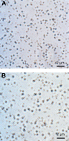
Optical microscopic images (600×). Notes: (A) Fe3O4-PLGA NPs and (B) Fe3O4-PLGA-cRGD NPs. The NPs had a regular shape and were relatively uniform in size. Abbreviations: cRGD, cyclic Arg-Gly-Asp; NPs, nanoparticles; PLGA, poly(lactic-co-glycolic acid).
Figure 3
Figure 3
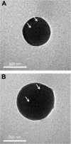
Transmission electron microscopic images. Notes: (A) Fe3O4-PLGA NPs and (B) Fe3O4-PLGA-cRGD nanoparticles. The iron oxide particles (white arrows) exhibited a relatively uniform distribution on the nanosized, spherical shell. Abbreviations: cRGD, cyclic Arg-Gly-Asp; PLGA, poly(lactic-co-glycolic acid).
Figure 4
Figure 4
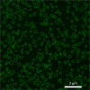
Laser scanning confocal microscopic image of the Fe3O4-PLGA-cRGD NPs. Note: Annular green fluorescence was observed around NPs, but almost no fluorescence was detected in the inner aqueous phase. Abbreviations: cRGD, cyclic Arg-Gly-Asp; NPs, nanoparticles; PLGA, poly(lactic-co-glycolic acid).
Figure 5
Figure 5
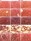
Pathologic hematoxylin and eosin staining (AE) of the heart, liver, spleen, lung, and kidney. (A1–E1) before NP injection, (A2–E2) after injection of Fe3O4-PLGA NPs, and (A3–E3) after injection of Fe3O4-PLGA-cRGD NPs. There was no evidence of inflammatory reactions or necrosis in the two groups, and no abnormal cell morphology was found. The magnification is 400×. Abbreviations: cRGD, cyclic Arg-Gly-Asp; NPs, nanoparticles; PLGA, poly(lactic-co-glycolic acid).
Figure 6
Figure 6
MRA, magnetic resonance imaging T2WI axial images, and pathologic H&E staining of the abdominal aorta. Notes: (A) Before induction of the thrombosis model, (B) post-induction, and (C) H&E staining of a section of the model. MRA revealed vessel lumen narrowing after the FeCl3-induced model was successfully constructed, and the signal in the abdominal aorta changed from low to high on the T2WI axial image. H&E staining confirmed that the mural thrombus was mixed. The magnification is 100×. Abbreviations: H&E, hematoxylin and eosin; MRA, magnetic resonance angiography; T2WI, T2-weighted imaging.
Figure 7
Figure 7
Hematoxylin and eosin staining of frozen sections of the ex vivo thrombus after injection of the NPs. Notes: (A) Fe3O4-PLGA NPs and (B) Fe3O4-PLGA-cRGD NPs. Many of the Fe3O4-PLGA-cRGD NPs gathered on the surface of the thrombus (black arrows), and no NPs were found on the surface of the thrombus in the sample with Fe3O4-PLGA NPs. The magnification is 200×. Abbreviations: cRGD, cyclic Arg-Gly-Asp; NPs, nanoparticles; PLGA, poly(lactic-co-glycolic acid).
Figure 8
Figure 8
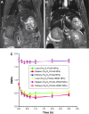
MRI T2WI images of the abdomen and graph of SNRs. Notes: (A) Before injection of the Fe3O4-PLGA-cRGD NPs. (B) After injection of the Fe3O4-PLGA-cRGD NPs at 30 min. (C) SNRs curves of the liver, spleen, and kidneys before and after the injection of Fe3O4-PLGA and Fe3O4-PLGA-cRGD NPs. MRI T2WI images of the abdomen demonstrated that the signals in the liver and spleen decreased significantly after NP injection. The SNRs of both the liver and spleen decreased significantly after NP injection as well, and the SNRs of the kidney decreased slightly. Abbreviations: cRGD, cyclic Arg-Gly-Asp; MRI, magnetic resonance imaging; NPs, nanoparticles; PLGA, poly(lactic-co-glycolic acid); SNRs, signal-to-noise ratios; T2WI, T2-weighted imaging.
Figure 9
Figure 9
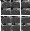
Magnetic resonance imaging T2WI images of the in vivo thrombosis before and after the injection of NPs. Notes: (A) Fe3O4-PLGA NPs and (B) Fe3O4-PLGA-cRGD NPs. Ten minutes after injection, the hyperintense signal of the abdominal aorta began to decrease and the signal of the peripheral zones of the abdominal aorta first obviously decreased and then gradually increased until 50 min in the Fe3O4-PLGA-cRGD NP group. There was no significant decrease in the signal of the abdominal aorta before or after injection in the Fe3O4-PLGA NP group. Abbreviations: cRGD, cyclic Arg-Gly-Asp; NPs, nanoparticles; PLGA, poly(lactic-co-glycolic acid); T2WI, T2-weighted imaging.
Figure 10
Figure 10
Hematoxylin and eosin staining of frozen sections of thrombi after injection with the NPs after in vivo magnetic resonance imaging scanning was finished. Notes: (A) Fe3O4-PLGA NPs and (B) Fe3O4-PLGA-cRGD NPs. A few of the Fe3O4-PLGA-cRGD NPs gathered on the surface of the thrombus, and many NPs were found under vascular endothelial cells (black arrows). No NPs were found in the sample injected with Fe3O4-PLGA NPs. These images are magnified ×200. Abbreviations: cRGD, cyclic Arg-Gly-Asp; NPs, nanoparticles; PLGA, poly(lactic-co-glycolic acid).

Similar articles

Cited by

References

    1. Whinna HC. Overview of murine thrombosis models. Thromb Res. 2008;122(Suppl 1):S64–S69. - PubMed
    1. Bender YY, Pfeifer A, Ebersberger HU, et al. Molecular cardiovascular magnetic resonance: current status and future prospects. Curr Cardiol Rep. 2016;18(5):47. - PubMed
    1. Previtali E, Bucciarelli P, Passamonti SM, Martinelli I. Risk factors for venous and arterial thrombosis. Blood Transfus. 2011;9(2):120–138. - PMC - PubMed
    1. Wu W, Wang Y, Shen S, et al. In vivo ultrasound molecular imaging of inflammatory thrombosis in arteries with cyclic Arg-Gly-Asp-modified microbubbles targeted to glycoprotein IIb/IIIa. Invest Radiol. 2013;48(11):803–812. - PubMed
    1. Wang X, Gkanatsas Y, Palasubramaniam J, et al. Thrombus-targeted theranostic microbubbles: a new technology towards concurrent rapid ultrasound diagnosis and bleeding-free fibrinolytic treatment of thrombosis. Theranostics. 2016;6(5):726–738. - PMC - PubMed