

Antitumor effects of oncolytic herpes simplex virus type 2 against colorectal cancer in vitro and in vivo
- PMID: 28223815
- PMCID: PMC5308569
- DOI: 10.2147/TCRM.S128575
Antitumor effects of oncolytic herpes simplex virus type 2 against colorectal cancer in vitro and in vivo
Abstract
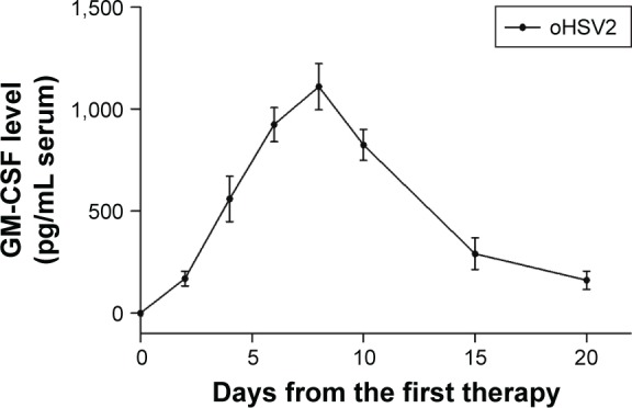
Background: The incidence of colorectal cancer (CRC) is on the rise. Furthermore, late-stage diagnoses and limited efficacious treatment options make CRC a complex clinical challenge. Therefore, a new therapeutic regimen with a completely novel therapeutic mechanism is necessary for CRC. In the present study, the therapeutic efficacy of oncolytic herpes simplex virus type 2 (oHSV2) in CRC was assessed in vitro and in vivo. oHSV2 is an oncolytic agent derived from herpes simplex virus type 2 that encodes granulocyte-macrophage colony-stimulating factor.
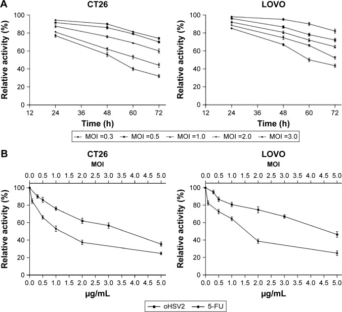
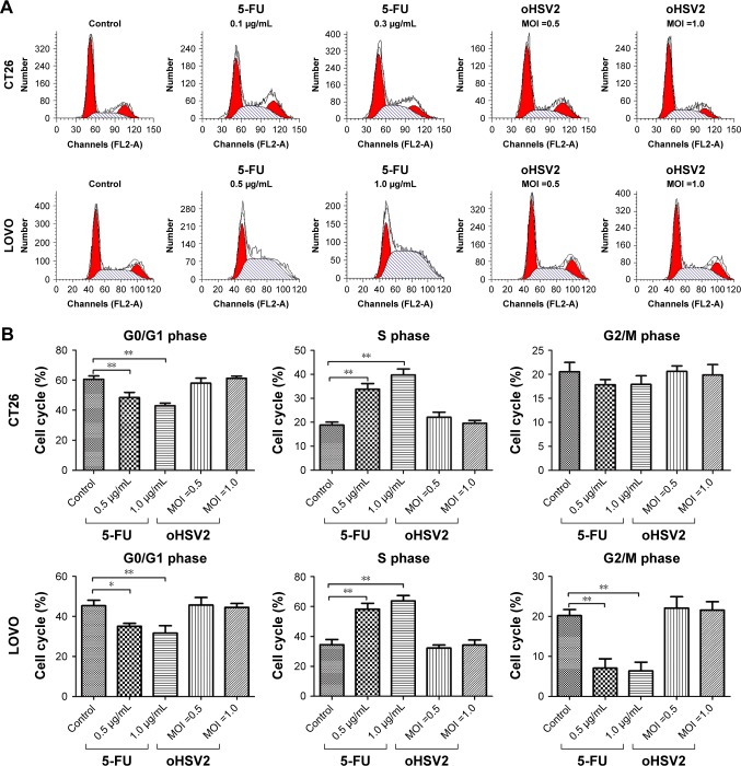
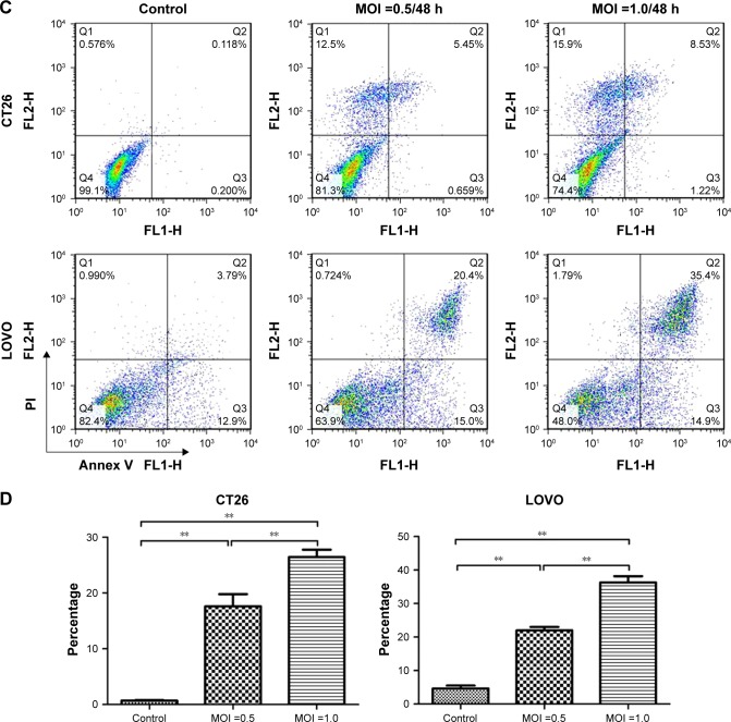
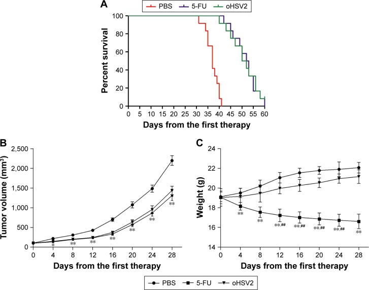
Materials and methods: We investigated the cytopathic effects of oHSV2 in CRC cell lines using the MTT assay. Then, cell cycle progression and apoptosis of oHSV2 were examined by flow cytometry. We generated a model of CRC with mouse CRC cell CT26 in BALB/c mice. The antitumor effects and adaptive immune response of oHSV2 were assessed in tumor-bearing mice. The therapeutic efficacy of oHSV2 was compared with the traditional chemotherapeutic agent, 5-fluorouracil.
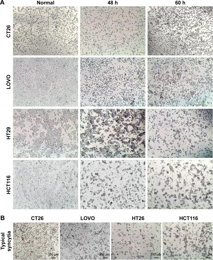
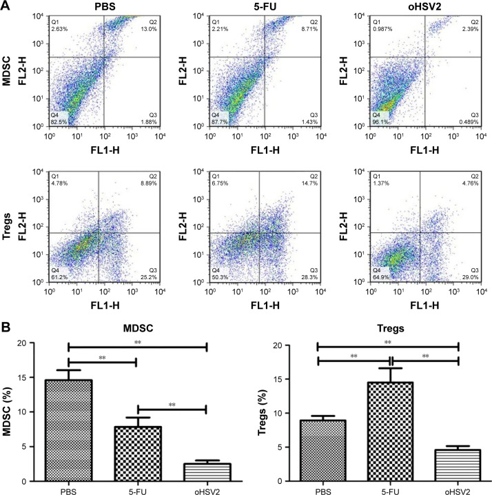
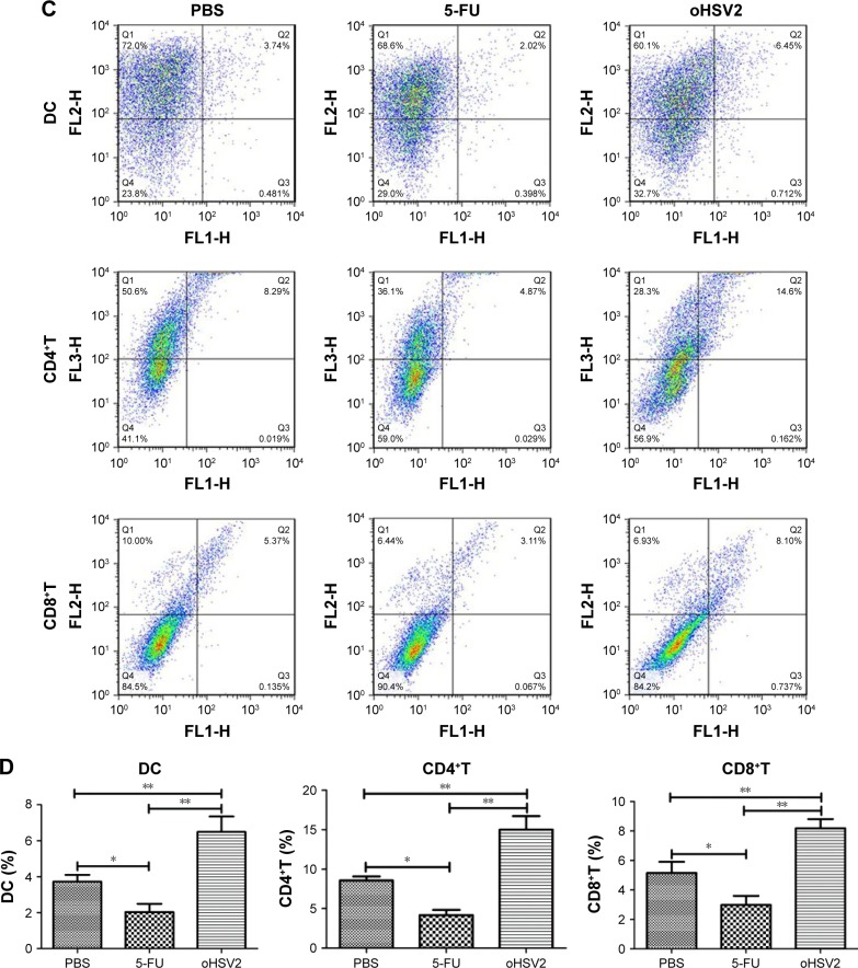
Results: The in vitro data showed that oHSV2 infected the CRC cell lines successfully and that the tumor cells formed a significant number of syncytiae postinfection. The oHSV2 killed cancer cells independent of the cell cycle and mainly caused tumor cells necrosis. The in vivo results showed that oHSV2 significantly inhibited tumor growth and prolonged survival of tumor-bearing mice without weight loss. With virus replication, oHSV2 not only resulted in a reduction of myeloid-derived suppressor cells and regulatory T cells in the spleen, but also increased the number of mature dendritic cells in tumor-draining lymph nodes and the effective CD4+T and CD8+T-cells in the tumor microenvironment.
Conclusion: Our study provides the first evidence that oHSV2 induces cell death in CRC in vitro and in vivo. These findings indicate that oHSV2 is an effective therapeutic cancer candidate that causes an oncolytic effect and recruits adaptive immune responses for an enhanced therapeutic impact, thus providing a potential therapeutic tool for treatment of CRC.
Keywords: colorectal cancer; gene therapy; granulocyte; herpes simplex virus type 2; immunotherapy; macrophage colony-stimulating factor; oncolytic virus.
Conflict of interest statement
Disclosure The authors report no conflicts of interest in this work.
Figures
Similar articles
-
Inhibition of colorectal cancer liver metastasis in BALB/c mice following intratumoral injection of oncolytic herpes simplex virus type 2 for the induction of specific antitumor immunity.Oncol Lett. 2019 Jan;17(1):815-822. doi: 10.3892/ol.2018.9720. Epub 2018 Nov 16. Oncol Lett. 2019. PMID: 30655834 Free PMC article.
-
Oncolytic virus oHSV2 combined with PD-1/PD-L1 inhibitors exert antitumor activity by mediating CD4 + T and CD8 + T cell infiltration in the lymphoma tumor microenvironment.Autoimmunity. 2023 Dec;56(1):2259126. doi: 10.1080/08916934.2023.2259126. Epub 2023 Sep 22. Autoimmunity. 2023. PMID: 37736847
-
A novel oncolytic herpes simplex virus type 2 has potent anti-tumor activity.PLoS One. 2014 Mar 26;9(3):e93103. doi: 10.1371/journal.pone.0093103. eCollection 2014. PLoS One. 2014. PMID: 24671154 Free PMC article.
-
Oncolytic herpes simplex virus and immunotherapy.BMC Immunol. 2018 Dec 18;19(1):40. doi: 10.1186/s12865-018-0281-9. BMC Immunol. 2018. PMID: 30563466 Free PMC article. Review.
-
Presage of oncolytic virotherapy for oral cancer with herpes simplex virus.Jpn Dent Sci Rev. 2017 May;53(2):53-60. doi: 10.1016/j.jdsr.2016.10.001. Epub 2016 Nov 5. Jpn Dent Sci Rev. 2017. PMID: 28479936 Free PMC article. Review.
Cited by
-
Inhibition of colorectal cancer liver metastasis in BALB/c mice following intratumoral injection of oncolytic herpes simplex virus type 2 for the induction of specific antitumor immunity.Oncol Lett. 2019 Jan;17(1):815-822. doi: 10.3892/ol.2018.9720. Epub 2018 Nov 16. Oncol Lett. 2019. PMID: 30655834 Free PMC article.
-
IL-24-Armed Oncolytic Vaccinia Virus Exerts Potent Antitumor Effects via Multiple Pathways in Colorectal Cancer.Oncol Res. 2021 Mar 16;28(6):579-590. doi: 10.3727/096504020X15942028641011. Epub 2020 Jul 8. Oncol Res. 2021. PMID: 32641200 Free PMC article.
-
Oncolytic virus-based hepatocellular carcinoma treatment: Current status, intravenous delivery strategies, and emerging combination therapeutic solutions.Asian J Pharm Sci. 2023 Jan;18(1):100771. doi: 10.1016/j.ajps.2022.100771. Epub 2022 Dec 29. Asian J Pharm Sci. 2023. PMID: 36896445 Free PMC article. Review.
-
Oncolytic Herpes Simplex Viral Therapy: A Stride toward Selective Targeting of Cancer Cells.Front Pharmacol. 2017 May 16;8:270. doi: 10.3389/fphar.2017.00270. eCollection 2017. Front Pharmacol. 2017. PMID: 28559846 Free PMC article. Review.
-
Immuno-Oncolytic Viruses: Emerging Options in the Treatment of Colorectal Cancer.Mol Diagn Ther. 2021 May;25(3):301-313. doi: 10.1007/s40291-021-00517-7. Epub 2021 Mar 12. Mol Diagn Ther. 2021. PMID: 33713031 Review.
References
-
- Siegel R, Naishadham D, Jemal A. Cancer statistics, 2013. CA Cancer J Clin. 2013;63:11–30. - PubMed
-
- Cunningham D, Atkin W, Lenz HJ, et al. Colorectal cancer. Lancet. 2010;375:1030–1047. - PubMed
-
- Ferlay J, Shin HR, Bray F, Forman D, Mathers C, Parkin DM. Estimates of worldwide burden of cancer in 2008: GLOBOCAN 2008. Int J Cancer. 2010;127:2893–2917. - PubMed
-
- Amin M, Lockhart AC. The potential role of immunotherapy to treat colorectal cancer. Expert Opin Investig Drugs. 2015;24(3):329–344. - PubMed
LinkOut - more resources
Full Text Sources
Other Literature Sources
Research Materials
