

Case Reports
Early Diagnosis of Intrahepatic Pseudoaneurysm during Radiofrequency Ablation using Contrast-Enhanced Ultrasound
Affiliations
- PMID: 28223887
- PMCID: PMC5295758
Item in Clipboard
Case Reports
Early Diagnosis of Intrahepatic Pseudoaneurysm during Radiofrequency Ablation using Contrast-Enhanced Ultrasound
Malays J Med Sci.
2015 Nov.
Abstract
Radiofrequency ablation is one of the more established forms of local treatment in patients with unresectable tumours, including colorectal hepatic metastases. Complications associated with this method of intervention include thermal and mechanical injuries, including vascular insults resulting in haemorrhage or pseudoaneurysm formation. This is the first case demonstrating the detection of post-ablation acute pseudoaneurysm formation identified on the table using contrast-enhanced ultrasound (CEUS) and the subsequent successful management.
Keywords: ablation; hepatic; metastasis; pseudoaneurysm; radiofrequency.
Conflict of interest statement
Conflict of Interest None.
Figures
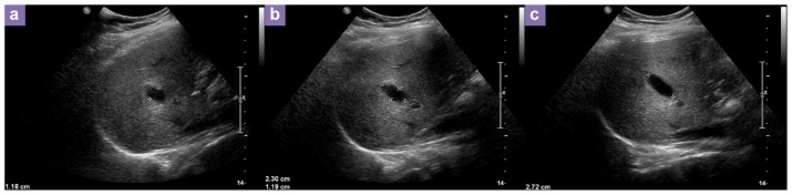
Sequential unenhanced ultrasound images show expanding pseudoaneurysm.
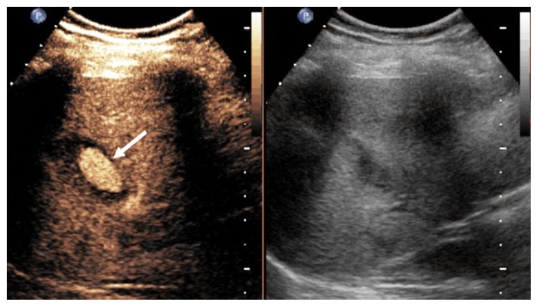
Contrast-enhanced ultrasound image shows microbubble contrast agent in the haematoma (arrow) confirming the pseudoaneurysm.
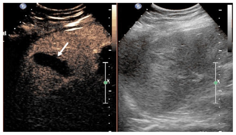
Post embolisation contrast-enhanced ultrasound image shows no contrast agent in the haematoma (arrow) confirming complete devascularization.

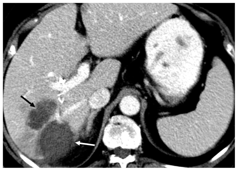
Computed tomography image shows focal enhancement in the pseudoaneurysm (black arrow) adjacent to the ablation zone (white arrow).
Computed tomography image shows no enhancement in the pseudoaneurysm (black arrow) adjacent to the ablation zone (white arrow).
Similar articles
-
Mycotic pseudoaneurysm of the hepatic artery as a complication of radiofrequency ablation of hepatic metastases.Radiol Case Rep. 2020 Oct 17;15(12):2663-2667. doi: 10.1016/j.radcr.2020.10.019. eCollection 2020 Dec. Radiol Case Rep. 2020. PMID: 33101562 Free PMC article.
-
Intrahepatic pseudoaneurysm after radiofrequency ablation of liver lesion.Int Surg. 2008 Nov-Dec;93(6):381-4. Int Surg. 2008. PMID: 20085050
-
Radiofrequency ablation for hepatocellular carcinoma: utility of conventional ultrasound and contrast-enhanced ultrasound in guiding and assessing early therapeutic response and short-term follow-up results.Ultrasound Med Biol. 2015 Sep;41(9):2400-11. doi: 10.1016/j.ultrasmedbio.2015.05.004. Epub 2015 Jun 6. Ultrasound Med Biol. 2015. PMID: 26055968 Clinical Trial.
-
Radiofrequency ablation for colorectal liver metastases.J Visc Surg. 2014 Apr;151 Suppl 1:S33-44. doi: 10.1016/j.jviscsurg.2013.12.005. Epub 2014 Feb 28. J Visc Surg. 2014. PMID: 24582728 Review.
-
Radiofrequency ablation of colorectal hepatic metastases.J Surg Oncol. 2010 Dec 15;102(8):978-87. doi: 10.1002/jso.21658. J Surg Oncol. 2010. PMID: 21166002 Review.
Cited by
-
Successful endovascular embolization of a giant splenic artery pseudoaneurysm secondary to a huge pancreatic pseudocyst with concomitant spleen invasion.Pol J Radiol. 2021 Aug 18;86:e489-e495. doi: 10.5114/pjr.2021.108876. eCollection 2021. Pol J Radiol. 2021. PMID: 34567295 Free PMC article.
References
Publication types
LinkOut - more resources
Full Text Sources