. 2017 May;25(5):591-599.
doi: 10.1038/ejhg.2017.9. Epub 2017 Feb 22.

Diagnostic exome sequencing in 266 Dutch patients with visual impairment

Affiliations

Diagnostic exome sequencing in 266 Dutch patients with visual impairment

Lonneke Haer-Wigman et al. Eur J Hum Genet. 2017 May.

Abstract

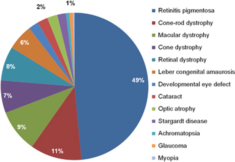
Inherited eye disorders have a large clinical and genetic heterogeneity, which makes genetic diagnosis cumbersome. An exome-sequencing approach was developed in which data analysis was divided into two steps: the vision gene panel and exome analysis. In the vision gene panel analysis, variants in genes known to cause inherited eye disorders were assessed for pathogenicity. If no causative variants were detected and when the patient consented, the entire exome data was analyzed. A total of 266 Dutch patients with different types of inherited eye disorders, including inherited retinal dystrophies, cataract, developmental eye disorders and optic atrophy, were investigated. In the vision gene panel analysis (likely), causative variants were detected in 49% and in the exome analysis in an additional 2% of the patients. The highest detection rate of (likely) causative variants was in patients with inherited retinal dystrophies, for instance a yield of 63% in patients with retinitis pigmentosa. In patients with developmental eye defects, cataract and optic atrophy, the detection rate was 50, 33 and 17%, respectively. An exome-sequencing approach enables a genetic diagnosis in patients with different types of inherited eye disorders using one test. The exome approach has the same detection rate as targeted panel sequencing tests, but offers a number of advantages. For instance, the vision gene panel can be frequently and easily updated with additional (novel) eye disorder genes. Determination of the genetic diagnosis improved the clinical diagnosis, regarding the assessment of the inheritance pattern as well as future disease perspective.


Conflict of interest statement

The authors declare no conflict of interest.

Figures

Figure 1
Figure 1
Clinical details of the 266 Dutch patients with visual impairment. In our cohort of patients with visual impairment 13 different (sub)types of inherited eye disorders were clinically diagnosed. Most cases had an IRD, including 129 patients (49%) with retinitis pigmentosa, 44 patients (16%) with cone or macular dystrophy, 30 patients (11%) with cone–rod dystrophy and 15 patients (6%) with Leber congenital amaurosis. Furthermore, six patients with cataract, six patients with developmental eye disorders, including patients with aniridia, coloboma, microcornea and micro- or nanopthalmos and six patients with optic atrophy were included. The group of retinal dystrophies contained unspecified retinal dystrophies (n=20) and rare forms of retinal dystrophies, including patients with benign concentric annular macular dystrophy, bradyopsia, congenital stationary night blindness, cystoid macular dystrophy, enhanced S-cone syndrome, foveal hypoplasia or night blindness with retinal detachment.
Figure 2
Figure 2
Mean percentage base pairs covered by at least 20 × of the vision panel genes. For each of the 298 vision panel genes, the mean percentage of base pairs covered by at least 20 × of the targeted gene region was calculated for all 48 samples on the SOLiD and for all 218 cases run on the HiSeq platform. On both platforms, most genes had a mean percentage of base pairs covered by at least 20 × in the range of 91 till 100%. However, if this range is further dissected, the SOLiD platform had most genes in the range of 91 till 95%, while for the HiSeq platform this was in the range of 96 till 100%. Furthermore, the HiSeq platform had only three genes with a mean percentage of base pairs covered by at least 20 × below 70%, while for the SOLiD platform this was the case in 65 genes.
Figure 3
Figure 3
Vision gene panel revisions and genes carrying variants in the vision gene panel analysis. (a) The genes present in the vision gene panel were grouped into seven main types of inherited visual impairment. In the 3-year time period of this study, the vision gene panel, used in the first step of analysis, was updated three times. The initial version of the panel consisted mostly of genes known to cause syndromic and/or non-syndromic IRD. In the second update also genes known to cause syndromic and/or non-syndromic cataract or developmental eye defects were added. Furthermore, in each update, recently published inherited eye disease genes were added. (b) In the vision gene panel analysis (likely) causative variants were detected in 56 different inherited eye disease genes. USH2A, EYS, ABCA4 and RPGR carried most often the causative variant and taken together they are the responsible causative gene in more than a quarter of the cases in whom a genetic cause was detected. The other genes were implicated to cause disease in 4% or less of the cases.
Figure 4
Figure 4
Flow-scheme of genetic tests that should be performed in patients with inherited eye disorders. In our exome approach, some regions of genes associated with inherited eye disorders have consistently low or no coverage. For instance, regions with high homology, GC-rich regions and repetitive regions are difficult to enrich. Moreover, deep-intronic and small copy number variations are not detected via an exome-sequencing approach. To provide the best care, a diagnostic flow-scheme specific for our exome approach was developed, to provide an overview in which types of inherited eye disorders a genetic test should be performed as a pre-screen or after a negative exome result. In patients with Leber congenital amaurosis, Stargardt disease, X-linked retinitis pigmentosa, blue cone monochromacy, Bornholm eye disease and albinism, a genetic pre-screen of one or two specific genes is indicated. In simplex male patients with retinitis pigmentosa or cone–rod dystrophy and in patients with X-linked cone–rod dystrophy, a genetic follow-up test of one or two specific genes is indicated after a negative exome result. In patients in whom a single heterozygous pathogenic variant is detected in a gene associated with autosomal recessive inheritance, it is indicated to test the non-covered regions of that gene.

References

    1. Kocur I, Resnikoff S: Visual impairment and blindness in Europe and their prevention. Br J Ophthalmol 2002; 86: 716–722. - PMC - PubMed
    1. Finger RP, Fimmers R, Holz FG, Scholl HP: Incidence of blindness and severe visual impairment in Germany: projections for 2030. Invest Ophthalmol Vis Sci 2011; 52: 4381–4389. - PubMed
    1. Ayuso C, Millan JM: Retinitis pigmentosa and allied conditions today: a paradigm of translational research. Genome Med 2010; 2: 34. - PMC - PubMed
    1. Sahel JA, Marazova K, Audo I: Clinical characteristics and current therapies for inherited retinal degenerations. Cold Spring Harb Perspect Med 2015; 5: a017111. - PMC - PubMed
    1. Rahi JS, Dezateux C, , British Congenital Cataract Interest G: Measuring and interpreting the incidence of congenital ocular anomalies: lessons from a national study of congenital cataract in the UK. Invest Ophthalmol Vis Sci 2001; 42: 1444–1448. - PubMed