. 2017 Feb 22:7:42940.
doi: 10.1038/srep42940.

Characterisation of urinary WFDC12 in small nocturnal basal primates, mouse lemurs (Microcebus spp.)

Affiliations

Characterisation of urinary WFDC12 in small nocturnal basal primates, mouse lemurs (Microcebus spp.)

Jennifer Unsworth et al. Sci Rep. .

Abstract

Mouse lemurs are basal primates that rely on chemo- and acoustic signalling for social interactions in their dispersed social systems. We examined the urinary protein content of two mouse lemurs species, within and outside the breeding season, to assess candidates used in species discrimination, reproductive or competitive communication. Urine from Microcebus murinus and Microcebus lehilahytsara contain a predominant 10 kDa protein, expressed in both species by some, but not all, males during the breeding season, but at very low levels by females. Mass spectrometry of the intact proteins confirmed the protein mass and revealed a 30 Da mass difference between proteins from the two species. Tandem mass spectrometry after digestion with three proteases and sequencing de novo defined the complete protein sequence and located an Ala/Thr difference between the two species that explained the 30 Da mass difference. The protein (mature form: 87 amino acids) is an atypical member of the whey acidic protein family (WFDC12). Seasonal excretion of this protein, species difference and male-specific expression during the breeding season suggest that it may have a function in intra- and/or intersexual chemical signalling in the context of reproduction, and could be a cue for sexual selection and species recognition.


Conflict of interest statement

The authors declare no competing financial interests.

Figures

Figure 1
Figure 1. Protein content of mouse lemur urine.
A group of 116 male and female mouse lemur (M. murinus and M. lehilahytsara) urine samples were analysed by SDS-PAGE (panel a, samples from several individuals, males: M1 to M8, females F1 to F9). Total urinary protein concentration generally increased with urinary creatinine, which provides a measure of urine dilution (panel b, symbols and colours represent donor sex and season). Some males in the breeding season had much higher levels of urinary protein than explained by urine dilution. The urinary protein output (expressed as μg protein/μg creatinine, averaged over replicate samples from the same individual and season) was higher in the breeding than in the non-breeding season among males, while there was no seasonal effect on female output (panel c, data are means ± sem, n indicates total number of individuals sampled). Multiple samples obtained from some individuals (10 males, 10 females) allowed urinary protein output to be compared between breeding and non-breeding season within the same individual (panel d).
Figure 2
Figure 2. Intact mass analysis of the mouse lemur urinary protein.
For urine samples from specific male mouse lemurs within the breeding season, the protein concentration was sufficiently high for analysis of the intact mass of the predominant protein by electrospray ionisation mass spectrometry. Urine samples were analysed for the two mouse lemur species, M. murinus and M. lehilahytsara, and representative deconvoluted mass spectra, showing the intact mass profiles are presented.
Figure 3
Figure 3. MALDI-TOF mass spectrometry of the mouse lemur urinary protein.
For some breeding season males, the predominant 10 kDa band was resolved by SDS-PAGE and the gel fragment was processed by reduction, carbamidomethylation and digestion with trypsin. The resultant peptides were analysed by MALDI-TOF mass spectrometry. Representative spectra are included for M. murinus (plotted in a positive direction, black) and M. lehilahytsara (plotted in a negative direction for spectral comparison, red). The two boxed peptides exhibited a 30 Da mass difference between the two species.
Figure 4
Figure 4. Complete sequencing strategy for the predominant 10 kDa protein.
Urine from M. murinus and M. lehilahytsara, containing high levels of the 10 kDa protein was reduced, carbamidomethylated and digested in solution with four different endoproteolytic enzymes (G: endopeptidase GluC, L: endopeptidase LysC, N:endopeptidase AspN and T: trypsin), the peptides were separated by reversed phase chromatography and sequenced de novo by high resolution tandem mass spectrometry. The top panel (a) summarises the sequencing strategy (the symbol ‘J’ is used to define ambiguity between isobaric leucine and isoleucine) The bottom panel (b) indicates the homology (MLP: mouse lemur protein sequenced in this study) with the protein inferred from the draft genome sequence for M. murinus, (MmWAP, number starting from 24 to accommodate the predicted signal peptide 1–23) assuming the alternate splice variant (see Fig. 5). The putative disulphide bond arrangement observed for other proteins containing the four disulphide core is superimposed on the new protein sequence. Additionally, the position of the single amino change between M. murinus and M. lehilahytsara and the site of methionine oxidation (for both species) are indicated. The masses of peptides observed by MALDI-TOF mass spectrometry are highlighted at the top of panel a, and the boxed peptide masses confirm the observations from MALDI-TOF mass spectra, where the observed peptides for M. murinus (m) or M. lehilahytsara (l) are identified.
Figure 5
Figure 5. Sequence comparison between mouse lemur and ring tailed lemur.
Panel (a) the predicted and observed mouse lemur sequences were aligned with the sequence from the ring tailed lemur (Lemur catta, Uniprot accession number A4K2S4). The position of an alternate splice junction in the mouse lemur sequence (http://www.ncbi.nlm.nih.gov/protein/829731644/) is indicated in red. The ambiguity between the isobaric pair leucine and isoleucine is indicated by the letter ‘J’. The dot plot (panel b) comparing the mouse lemur and ‘long form’ predicted ring tailed lemur sequences was generated using the dotPlot routine within the seqinr R package (http://seqinr.r-forge.r-project.org/) with a window size of five and scoring matches of three or more amino acids within the window. Panel (c) - Inferred exon/intron structure of WFDC12 in Microcebus murinus genomic sequence of WFDC12 from Ensembl record ENSMICG00000000986. The four exons from automated prediction are underlined. The three inferred actual exons that encode the WFDC12 protein sequenced in this study are in blue font with coloured background – the translation of these 3 spliced exons is shown as “M murinus pred” in panel b. Exons 1 and 2 and intron 1 are the same in both annotations. The predicted 5′ and 3′ splice sites for the newly defined intron 2 are circled and coloured red, with an AG 3′ splice site immediately upstream of exon 3, as expected.
Figure 6
Figure 6. The primate WFDC12 family.
All available primate WFDC12 proteins, were aligned using the CLC Sequence Viewer (www.clcbio.com), version 7.6.1. The eight fully conserved cysteine residues are coloured yellow in the conservation graph, in addition to the further 15 residues (blue) within the four disulphide domain that are conserved across all primates shown here.
Figure 7
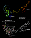
Figure 7. Homology modelling of the mouse lemur urinary protein.
The sequence of the mouse lemur protein from M. murinus was used to obtain a model structure using the Phyre2 server (http://www.sbg.bio.ic.ac.uk/~phyre2) using the ‘intensive’ modelling approach. The four disulphide core was modelled to a very level of confidence, but the C-terminal domain cannot be predicted with equivalent confidence.

References

    1. Wyatt T. D. Pheromones and animal behavior: chemical signals and signatures (Cambridge University Press, Cambridge, 2013).
    1. Liberles S. D. Mammalian pheromones. Annu Rev Physiol 76, 151–175 (2014). - PMC - PubMed
    1. Kaur A. W. et al.. Murine pheromone proteins constitute a context-dependent combinatorial code governing multiple social behaviors. Cell 157, 676–688 (2014). - PMC - PubMed
    1. Roberts S. A. et al.. Darcin: a male pheromone that stimulates female memory and sexual attraction to an individual male’s odour. BMC Biol 8, 75 (2010). - PMC - PubMed
    1. Roberts S. A., Davidson A. J., McLean L., Beynon R. J. & Hurst J. L. Pheromonal induction of spatial learning in mice. Science 338, 1462–1465 (2012). - PubMed

Publication types

LinkOut - more resources