. 2017 Feb 22:7:43158.
doi: 10.1038/srep43158.

Testicular Dnmt3 expression and global DNA methylation are down-regulated by gonadotropin releasing hormones in the ricefield eel Monopterus albus

Affiliations

Testicular Dnmt3 expression and global DNA methylation are down-regulated by gonadotropin releasing hormones in the ricefield eel Monopterus albus

Yize Zhang et al. Sci Rep. .

Abstract

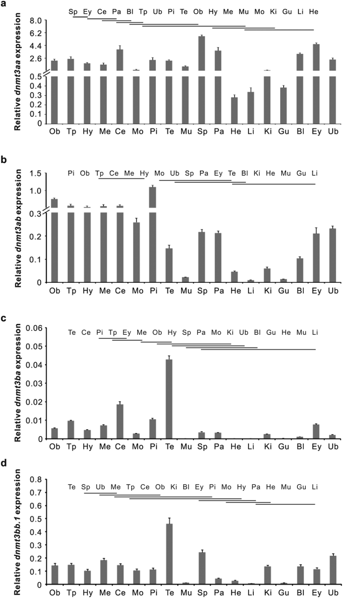
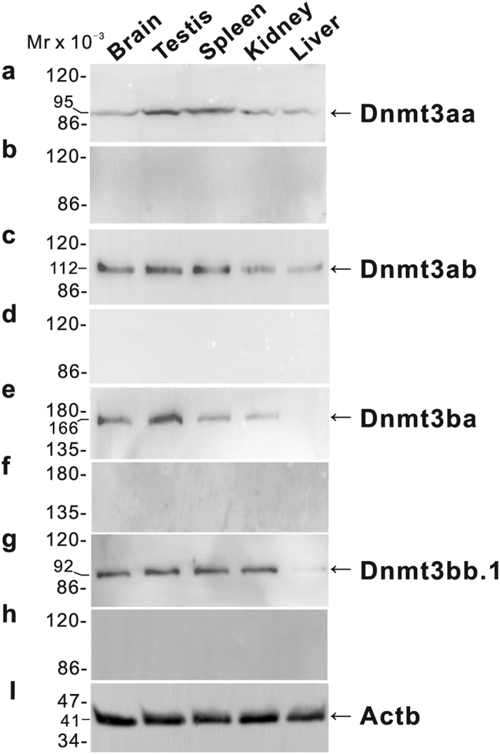
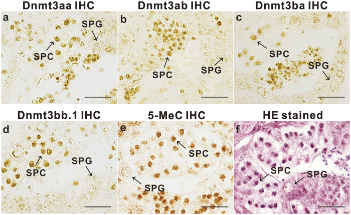
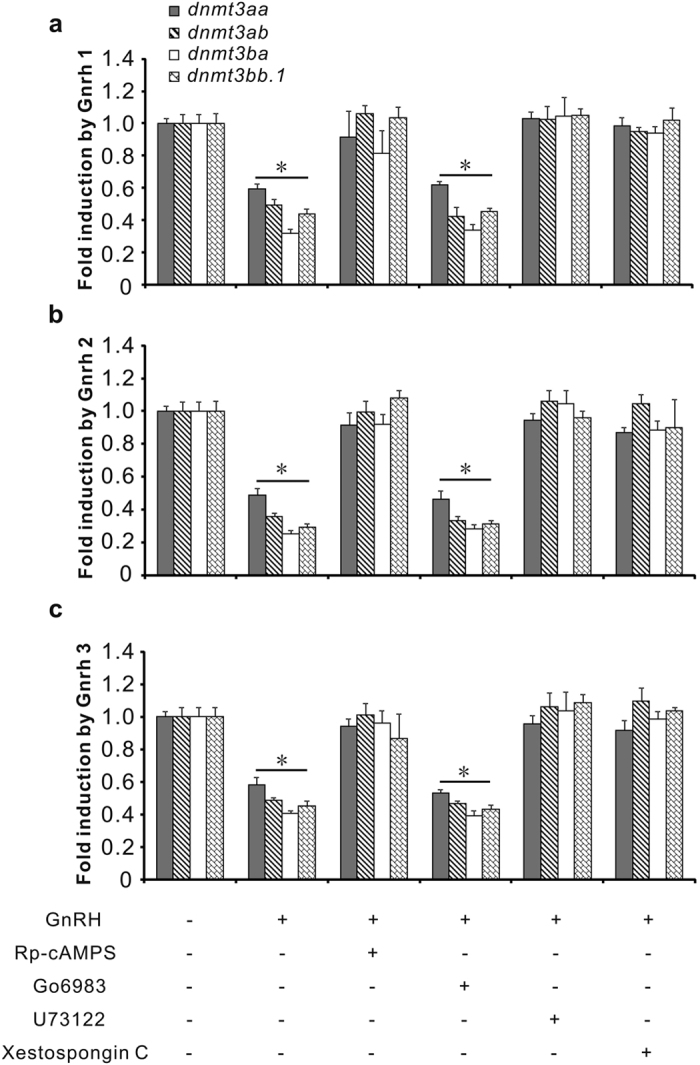
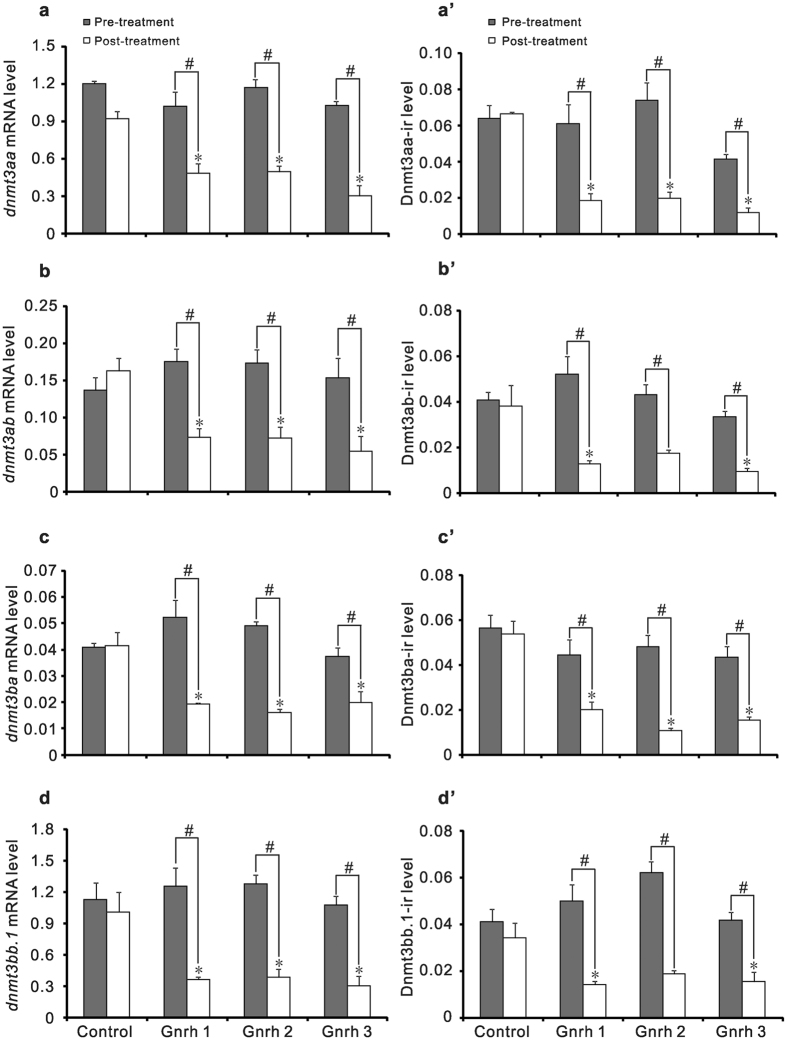
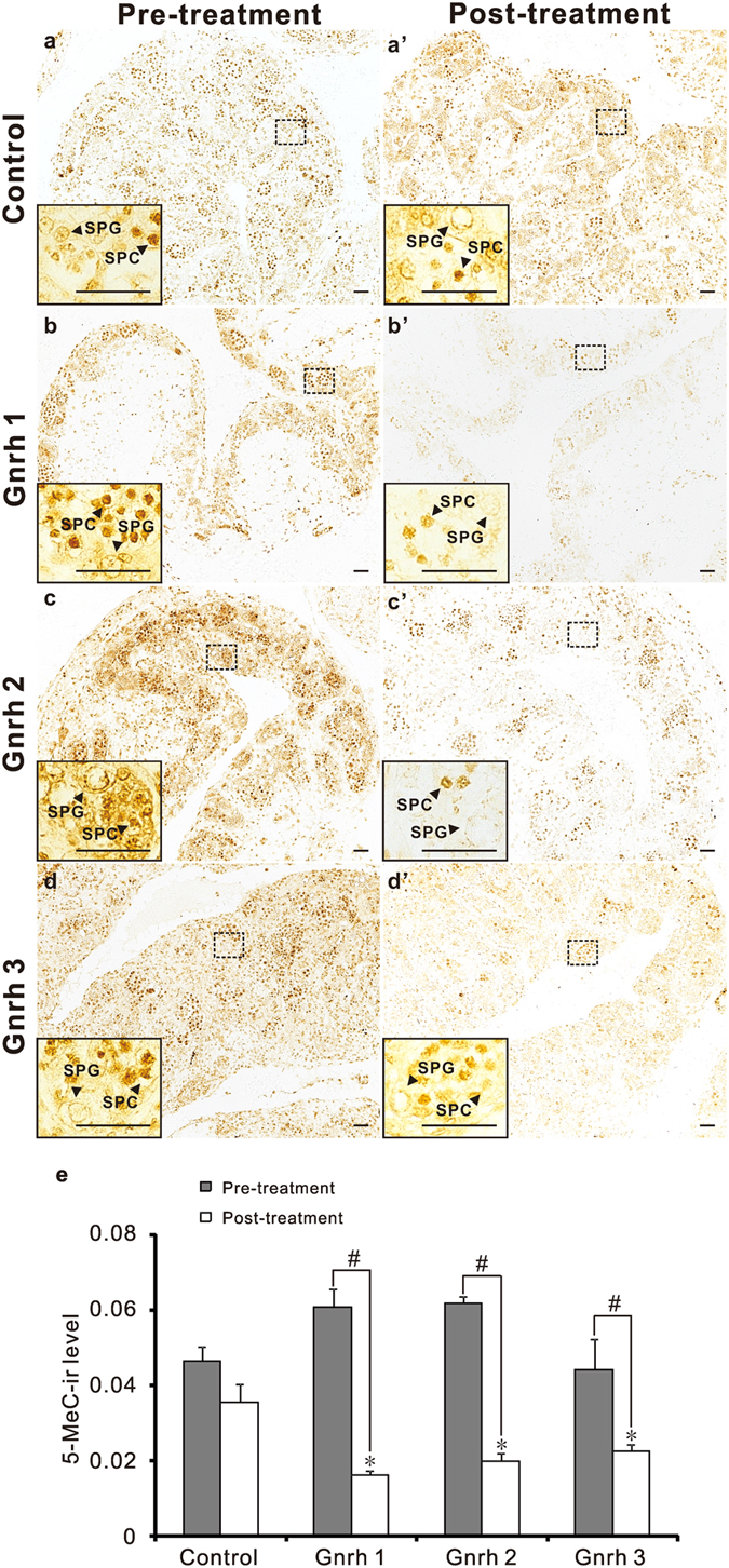
In vertebrates, DNA methyltransferase 3 (Dnmt3) homologues are responsible for de novo DNA methylation and play important roles in germ cell development. In the present study, four dnmt3 genes, dnmt3aa, dnmt3ab, dnmt3ba and dnmt3bb.1, were identified in ricefield eels. Real-time quantitative PCR analysis showed that all four dnmt3 mRNAs were detected broadly in tissues examined, with testicular expression at relatively high levels. In the testis, immunostaining for all four Dnmt3 forms was mainly localized to spermatocytes, which also contained highly methylated DNA. All three forms of Gonadotropin-releasing hormone (Gnrh) in the ricefield eel were shown to decrease the expression of dnmt3 genes in the in vitro incubated testicular fragments through cAMP and IP3/Ca2+ pathways. Moreover, in vivo treatment of male fish with three forms of Gnrh decreased significantly the testicular Dnmt3 expression at both mRNA and protein levels, and the global DNA methylation levels. These results suggest that the expression of Dnmt3 and global DNA methylation in the testis of ricefield eels are potentially down-regulated by Gnrh, and reveal a novel regulatory mechanism of testicular Dnmt3 expression in vertebrates.


Conflict of interest statement

The authors declare no competing financial interests.

Figures

Figure 1
Figure 1
Real-time quantitative PCR analysis of dnmt3aa (a), dnmt3ab (b), dnmt3ba (c) and dnmt3bb.1 (d) mRNA levels in tissues of male ricefield eels. The tissues analyzed are indicated below the corresponding bars. Bl: blood; Ce: cerebellum; Ey: eye; Gu: gut; He: heart; Hy: hypothalamus; Ki: kidney; Li: liver; Me: mesencephalon; Mo: medulla oblongata; Mu: muscle; Ob: olfactory bulb; Pa: pancreas; Pi: pituitary; Sp: spleen; Tp: telencephalon; Te: testis; Ub: urinary bladder. Each bar represents the mean of normalized expression levels ± SEM of 6 replicates. The results of statistical analysis were indicated at the top of each histogram, ranking from the high (left) to the low (right). The mRNA levels for tissues with common underscoring are not significantly different (P > 0.05).
Figure 2
Figure 2. Western blot analysis of Dnmt3 proteins in tissues of male ricefield eels.
The tissue homogenates from the brain, testis, spleen, kidney, and liver were separated on 8% SDS-PAGE gels, transferred to polyvinylidene difluoride membranes, and then immunoreacted with (a) anti-Dnmt3aa antiserum (1:1000); (b) anti-Dnmt3aa antiserum pre-adsorbed by excessive recombinant Dnmt3aa-N; (c) anti-Dnmt3ab antiserum (1:1000); (d) anti-Dnmt3ab antiserum pre-adsorbed by excessive recombinant Dnmt3ab-N; (e) antiDnmt3ba antiserum (1:1000); (f) anti-Dnmt3ba antiserum pre-adsorbed by excessive recombinant Dnmt3ba-N; (g) anti-Dnmt3bb.1 antiserum (1:1000); (h) anti-Dnmt3bb.1 antiserum pre-adsorbed by excessive recombinant Dnmt3bb.1-N; (i) mouse anti-Actb monoclonal antibody (1:500, 60008-1-Ig; ProteinTech Group, Inc. IL, USA). The secondary antibody was 1:5000 diluted horseradish peroxidase (HRP)-conjugated goat anti-mouse IgG H + L (115-035-003, Jackson ImmunoResearch Laboratories, Inc.). The blots were visualized using a BeyoECL Plus kit (Beyotime).
Figure 3
Figure 3. Cellular localization of immunoreactive Dnmt3 and global DNA methylation in testes of ricefield eels.
Immunostaining for Dnmt3aa (a), Dnmt3ab (b), Dnmt3ba (c), Dnmt3bb.1 (d), and 5-Methylcytosine (e) and hematoxylin-eosin staining (f) were performed on sections from the same testicular tissue. The antiserum against Dnmt3aa (1:200), Dnmt3ab (1:200), Dnmt3ba (1:200), Dnmt3bb.1 (1:200) or 5-MeC (1:200) was used as the primary antiserum. The secondary antibody was 1:500 diluted horseradish peroxidase (HRP)-conjugated goat anti-mouse IgG H + L (115-035-003, Jackson ImmunoResearch Laboratories, Inc.). The immunoreactive signals were visualized by DAB chromogen. SPC, spermatocyte; SPG, spermatogonium; Scale bar = 50 μm.
Figure 4
Figure 4
Effects of Gnrh 1, Gnrh 2 and Gnrh 3 on the expression of dnmt3aa (a), dnmt3ab (b), dnmt3ba (c) and dnmt3bb.1 (d) in the in vitro incubated testicular fragments of male ricefield eels. The testicular fragments were pre-incubated for 18 h before treating with Gnrh 1, Gnrh 2, or Gnrh 3 (10 and 100 nM) for 8 h. After treatment, mRNA levels of dnmt3aa, dnmt3ab, dnmt3ba, and dnmt3bb.1 in the testicular fragments were quantified with real-time quantitative PCR. Each bar represents the mean ± SEM of triplicates. The experiments were repeated three times, and similar results were obtained. Means marked with different letters are significantly different (P < 0.05).
Figure 5
Figure 5. Effects of inhibitors of intracellular signaling pathways on Gnrh down-regulation of dnmt3aa, dnmt3ab, dnmt3ba, and dnmt3bb.1 in the in vitro incubated testicular fragments of ricefield eels.
The testicular fragments were pre-incubated for 18 h before treating with 100 nM of Gnrh 1 (a), Gnrh 2 (b), and Gnrh 3 (c) in the presence or absence of inhibitors Rp-cAMPS (50 μM), Go6983 (10 μM), U73122 (10 μM) or Xestospongin C (1 μM) respectively for 8 h. After treatment, mRNA levels of dnmt3aa, dnmt3ab, dnmt3ba, and dnmt3bb.1 in the testicular fragments were quantified with real-time quantitative PCR and were presented as fold change relative to the vehicle control. Each bar represents mean ± SEM of 6 replicates. *P < 0.05 vs the vehicle control.
Figure 6
Figure 6
Effects of intraperitoneal injections of Gnrh 1, Gnrh 2 and Gnrh 3 on levels of dnmt3 mRNAs (a~d) and Dnmt3 proteins (a’~d’) in testes of ricefield eels. The male ricefield eels received intraperitoneal injections of Gnrh 1, Gnrh 2, or Gnrh 3 (0.1 μg/g body weight) twice a week for four weeks. After treatments, the mRNA levels of dnmt3aa (a), dnm3ab (b), dnmt3ba (c), and dnmt3bb.1 (d) in testes were quantified with real-time PCR. The immunoreactive Dnmt3aa (a’), Dnmt3ab (b’), Dnmt3ba (c’) and Dnmt3bb.1 (d’) levels in testes were analyzed by immunohistochemitry and quantified as described in Methods. Each bar represents mean ± SEM (n = 3~4). The experiments were repeated twice and similar results were obtained. *P < 0.05 vs the control; #P < 0.05 for the differences between the indicated groups.
Figure 7
Figure 7. Effects of intraperitoneal injections of Gnrh 1, Gnrh 2, or Gnrh 3 on immunoreactive 5-Methylcytosine (5-MeC) levels in testes of ricefield eels.
The male ricefield eels were first examined by biopsy and then received intraperitoneal injections of Gnrh 1, Gnrh 2, or Gnrh 3 (0.1 μg/g body weight) twice a week for four weeks. The 5-MeC level in the testis was analyzed with the immunohistochemistry as described in Methods, and immunoreactive signals were visualized by DAB chromogen. 5-MeC (1:200) antibody was used as the primary antibody. The secondary antibody was 1:500 diluted horseradish peroxidase (HRP)-conjugated goat anti-mouse IgG H + L (115-035-003, Jackson ImmunoResearch Laboratories, Inc.). The representative testicular sections of the same fish before (a~d) and after (a’~d’) treatments were shown, and the insets are the higher magnification of the boxed areas within each image, respectively. The immunoreactive 5-MeC levels are shown in panel e. SPC: spermatocyte; SPG: spermatogonium. Each bar in panel e represents the mean ± SEM (n = 3~4). The experiments were repeated twice and similar results were obtained. *P < 0.05 vs the control; #P < 0.05 for the differences between the indicated groups. Scale bar = 25 μm.

References

    1. Reik W., Dean W. & Walter J. Epigenetic reprogramming in mammalian development. Science 293, 1089–1093 (2001). - PubMed
    1. Goll M. G. & Bestor T. H. Eukaryotic cytosine methyltransferases. Annu Rev Biochem 74, 481–514 (2005). - PubMed
    1. Bird A. DNA methylation patterns and epigenetic memory. Genes Dev 16, 6–21 (2002). - PubMed
    1. Hermann A., Gowher H. & Jeltsch A. Biochemistry and biology of mammalian DNA methyltransferases. Cell Mol Life Sci 61, 2571–2587 (2004). - PMC - PubMed
    1. Okano M., Bell D. W., Haber D. A. & Li E. DNA Methyltransferases Dnmt3a and Dnmt3b are essential for de novo methylation and mammalian development. Cell 99, 247–257 (1999). - PubMed

Publication types

Substances