. 2017 Feb 22:7:43251.
doi: 10.1038/srep43251.

UVRAG Deficiency Exacerbates Doxorubicin-Induced Cardiotoxicity

Affiliations

UVRAG Deficiency Exacerbates Doxorubicin-Induced Cardiotoxicity

Lin An et al. Sci Rep. .

Abstract

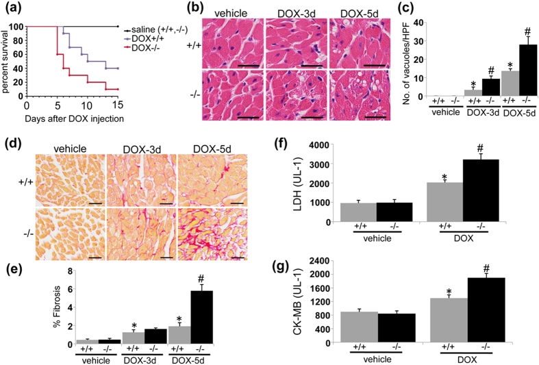
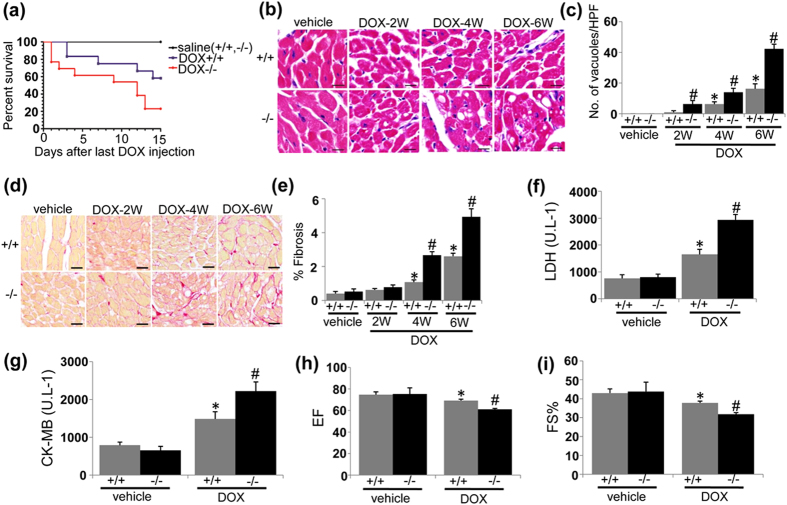
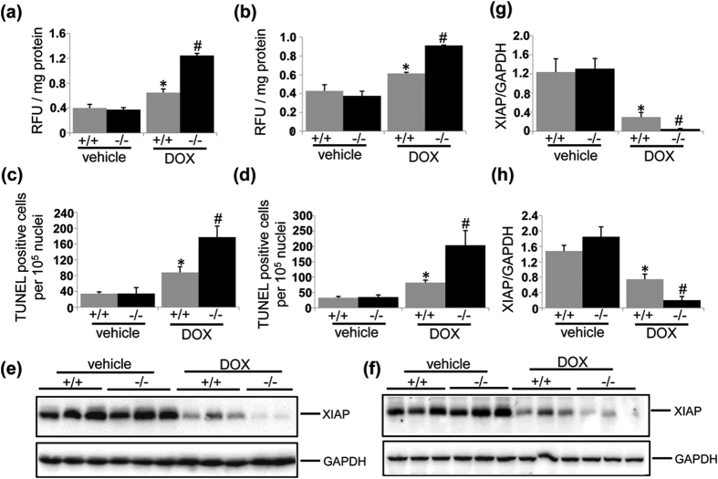
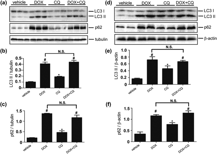
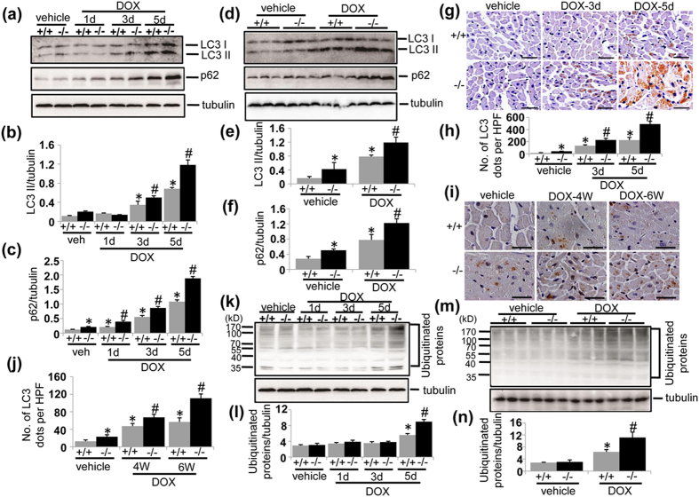
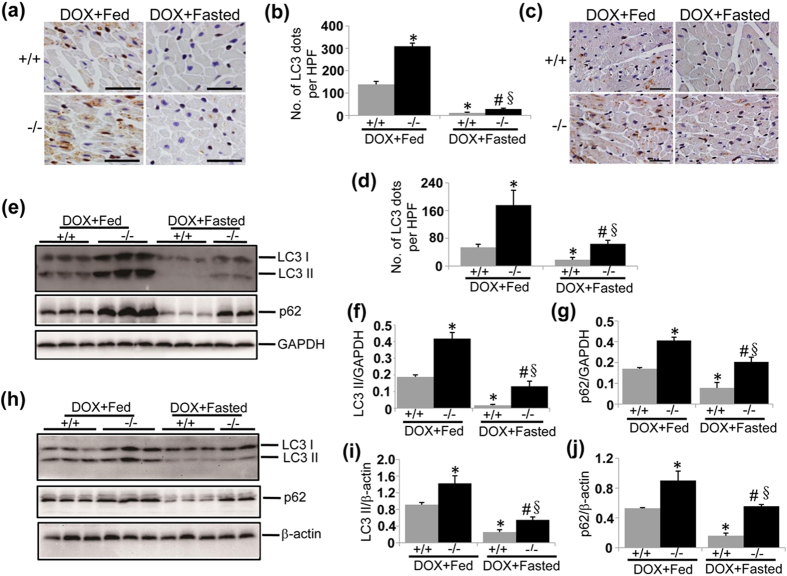
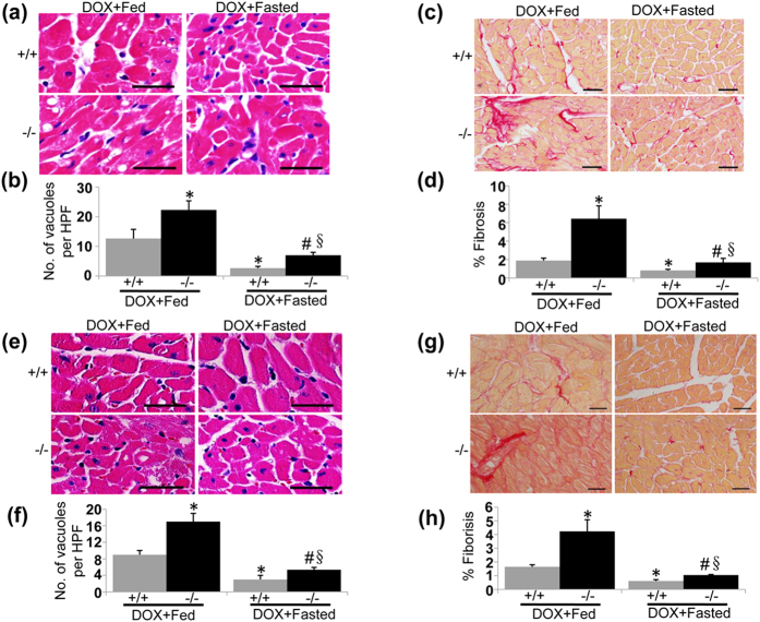
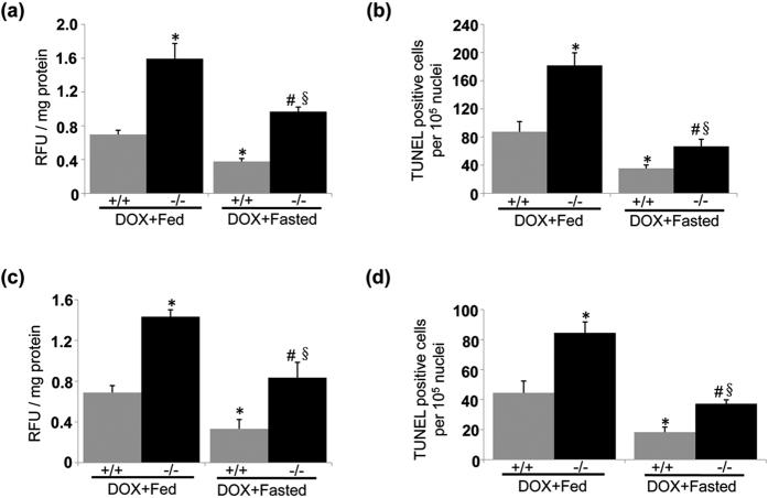
Doxorubicin (DOX) is an effective chemotherapeutic drug in the treatment of various types of cancers. However, its clinical application has been largely limited by potential development of cardiotoxicity. Previously we have shown that ultra-violet radiation resistance-associated gene (UVRAG), an autophagy-related protein, is essential for the maintenance of autophagic flux in the heart under physiological conditions. Here, we sought to determine the role of UVRAG-mediated autophagy in DOX-induced cardiotoxicity. Mouse models of acute or chronic DOX-induced cardiotoxicity were established. UVRAG deficiency exacerbated DOX-induced mortality and cardiotoxicity manifested by increased cytoplasmic vacuolization, enhanced collagen accumulation, elevated serum activities of lactate dehydrogenase and myocardial muscle creatine kinase, higher ROS levels, aggravated apoptosis and more depressed cardiac function. Autophagic flux was impaired in DOX-induced cardiotoxicity. UVRAG deficiency aggravated impaired autophagic flux in DOX-induced cardiotoxicity. Intermittent fasting restored autophagy and ameliorated pathological alterations of DOX-induced cardiotoxicity. Collectively, our data suggest that UVRAG deficiency exacerbates DOX-induced cardiotoxicity, at least in part, through aggravation of DOX-induced impaired autophagic flux. Intermittent fasting, which restores blunted autophagic flux and ameliorates pathology in the mouse models of DOX-induced cardiotoxicity, may be used as a potential preventive or therapeutic approach for DOX cardiotoxicity.


Conflict of interest statement

The authors declare no competing financial interests.

Figures

Figure 1
Figure 1. UVRAG deficiency exacerbates acute DOX-induced cardiotoxicity.
(a) Survival curves of WT and UVRAG-deficient mice after acute DOX treatment were created by Kaplan-Meier method (*P < 0.05 using the Log-rank test). n = 7 mice for saline+/+, n = 8 mice for saline−/−, n = 10 mice for DOX+/+, n = 10 mice for DOX−/−. (b) Representative H&E images of degenerative vacuoles in LVs on heart sections from WT and UVRAG-deficient mice 3 or 5 days after acute DOX or vehicle treatment. Scale bar: 40 μm. (c) Quantification of degenerative vacuoles in LVs in the experiments as illustrated in (b). n = 3 mice for each group. (d) Representative images of fibrosis stained with picrosirius red in LVs on heart sections from WT and UVRAG-deficient mice 3 or 5 days after acute DOX or vehicle treatment. Scale bar: 40 μm. (e) Quantification of fibrosis in LVs in the experiments as illustrated in (d). n = 3 mice for each group. (f) Serum LDH activity in WT and UVRAG-deficient mice 3 days after acute DOX or vehicle treatment. n = 4 mice for each group. (g) Serum CK-MB activity in WT and UVRAG-deficient mice 3 days after acute DOX or vehicle treatment. n = 4 mice for each group. *P < 0.05 vs. WT + vehicle, #P < 0.05 vs. WT + DOX.
Figure 2
Figure 2. UVRAG deficiency exacerbates chronic DOX-induced cardiotoxicity.
(a) Survival curves of WT and UVRAG-deficient mice after chronic DOX treatment were created by Kaplan-Meier method (*P < 0.05 using the Log-rank test). n = 6 mice for saline + / + , n = 6 mice for saline−/−, n = 12 mice for DOX + / + , n = 13 mice for DOX−/−. (b) Representative H&E images of degenerative vacuoles in LVs on heart sections from WT and UVRAG-deficient mice after chronic DOX or vehicle treatment over the indicated time. Scale bar: 40 μm. (c) Quantification of degenerative vacuoles in LVs in the experiments as illustrated in (b). n = 3–5 mice for each group. (d) Representative images of fibrosis stained with picrosirius red in LVs on heart sections from WT and UVRAG-deficient mice after chronic DOX or vehicle treatment over the indicated time. Scale bar: 40 μm. (e) Quantification of fibrosis in LVs in the experiments as illustrated in (d). n = 3–5 mice for each group. (f) Serum LDH activity in WT and UVRAG-deficient mice 2 weeks after chronic DOX or vehicle treatment. n = 4 mice for each group. (g) Serum CK-MB activity in WT and UVRAG-deficient mice 2 weeks after chronic DOX or vehicle treatment. n = 4 mice for each group. (h) Left ventricular EF of WT and UVRAG-deficient mice after chronic DOX or vehicle treatment. n = 4–5 mice for each group. (i) Left ventricular FS of WT and UVRAG-deficient mice after chronic DOX or vehicle treatment. n = 4–5 mice for each group. *P < 0.05 vs. WT + vehicle, #P < 0.05 vs. WT + DOX.
Figure 3
Figure 3. UVRAG deficiency increases ROS production and apoptosis in DOX-induced cardiotoxicity.
(a) ROS levels in LVs detected using DCFH-DA 5 days after acute DOX or vehicle treatment. n = 3 mice for each group. (b) ROS levels in LVs detected using DCFH-DA at 4 weeks of DOX or vehicle treatment in chronic DOX cardiotoxicity. n = 3 mice for each group. (c) Quantification of apoptotic cells 5 days after acute DOX or vehicle treatment. n = 3 mice for each group. (d) Quantification of apoptotic cells at 4 weeks of DOX or vehicle treatment. (e) Western blot analysis of XIAP in LVs from WT and UVRAG-deficient mice 3 days after acute DOX or vehicle treatment. (f) Quantification of XIAP as illustrated in panel (e). (g) Western blot analysis of XIAP in LVs from WT and UVRAG-deficient mice at 4 weeks of chronic DOX treatment or vehicle treatment. (h) Quantification of XIAP as illustrated in panel (g). n = 3 mice for each group. *P < 0.05 vs. WT + vehicle, #P < 0.05 vs. WT + DOX.
Figure 4
Figure 4. Autophagic flux is impaired in acute and chronic DOX-induced cardiotoxicity.
(a) Western blot detection of LC3 II and p62 protein abundance in LVs from mice 5 days after acute DOX treatment and further elicited by CQ treatment. (b) Quantification of LC3-II protein abundance as illustrated in panel (a). n = 3 mice for each group. (c) Quantification of p62 protein abundance as illustrated in panel (a). n = 3 mice for each group. (d) Western blot detection of LC3 II and p62 protein abundance in LVs from mice treated with DOX over 4 weeks and further elicited by CQ treatment. (e) Quantification of LC3-II as illustrated in panel (d). n = 3 mice for each group. (f) Quantification of p62 as illustrated in panel (d). n = 3 mice for each group. *P < 0.05 vs. vehicle. #P < 0.05 vs. CQ.
Figure 5
Figure 5. UVRAG deficiency aggravates impaired autophagy in DOX-induced cardiotoxicity.
(a) Western blot detection of LC3 II and p62 protein levels in LVs from WT and UVRAG-deficient mice at the time points as indicated in acute DOX-treated mice. (b) Quantification of LC3 II protein abundance in the experiments as illustrated in (a). n = 3–5 mice for each group. (c) Quantification of p62 protein abundance in the experiments as illustrated in (a). n = 3–5 mice for each group. (d) Western blot detection of LC3 II and p62 protein levels in LVs from WT and UVRAG-deficient mice at 4 weeks of DOX or vehicle treatment in chronic DOX-treated mice. (e) Quantification of LC3 II protein abundance in the experiments as illustrated in (d). n = 3 mice for each group. (f) Quantification of p62 protein abundance in the experiments as illustrated in (d). n = 3 mice for each group. (g) Representative images of LC3 immunohistochemistry in LVs on heart sections from WT and UVRAG-deficient mice after acute DOX or vehicle treatment over the indicated time. Scale bar: 40 μm. (h) Quantification of LC3-positive dots in LVs in the experiments as illustrated in (g). n = 3 mice for each group. (i) Representative images of LC3 immunohistochemistry in LVs on heart sections from WT and UVRAG-deficient mice after chronic DOX or vehicle treatment over the indicated time. Scale bar: 40 μm. (j) Quantification of LC3-positive dots in LVs in the experiments as illustrated in (i). n = 3 mice for each group. (k) Western blot detection of ubiquitinated proteins in LVs from WT and UVRAG-deficient mice at the time points as indicated in acute DOX-treated mice. (l) Quantification of protein ubiquitination in the experiments as illustrated in (k). n = 3 mice for each group. (m) Western blot detection of ubiquitinated proteins in LVs from WT and UVRAG-deficient mice at the time points as indicated in chronic DOX-treated mice. (n) Quantification of protein ubiquitination in the experiments as illustrated in (m). n = 3 mice for each group. *P < 0.05 vs. WT + vehicle. #P < 0.05 vs. WT + DOX.
Figure 6
Figure 6. Intermittent fasting restores autophagy in DOX-induced cardiotoxicity.
(a) Representative images of LC3 immunohistochemistry in LVs on heart sections from fed or fasted WT and UVRAG-deficient mice 5 days after acute DOX or vehicle treatment. Scale bar: 40 μm. (b) Quantification of LC3-positive dots in LVs in the experiments as illustrated in (a). n = 3 mice for each group. (c) Representative images of LC3 immunohistochemistry in LVs on heart sections from fasted WT and UVRAG-deficient mice at 4 weeks of DOX treatment in chronic cardiotoxicity. Scale bar: 40 μm. (d) Quantification of LC3-positive dots in LVs in the experiments as illustrated in (c). n = 3 mice for each group. (e) Western blot detection of LC3 and p62 in LVs from fed or fasted WT and UVRAG-deficient mice 5 days after acute DOX or vehicle treatment. (f) Quantification of LC3 II protein abundance in the experiments as illustrated in (e). n = 3 mice for each group. (g) Quantification of p62 protein abundance in the experiments as illustrated in (e). n = 3 mice for each group. (h) Western blot analysis of LC3 II and p62 in LVs from fed or fasted WT and UVRAG-deficient mice at 4 weeks of DOX treatment in chronic cardiotoxicity. (i) Quantification of LC3 II protein abundance in the experiments as illustrated in (h). n = 3 mice for each group. (j) Quantification of p62 protein abundance in the experiments as illustrated in (h). n = 3 mice for each gorup. *P < 0.05 vs. WT + DOX + Fed, #P < 0.05 vs. UVRAG−/− + DOX + Fed, §P < 0.05 vs. WT + DOX + Fasted.
Figure 7
Figure 7. Intermittent fasting ameliorates pathology of DOX-induced cardiotoxicity.
(a) Representative H&E images of degenerative vacuoles in LVs on heart sections from fed or fasted WT and UVRAG-deficient mice 5 days after acute DOX or vehicle treatment. Scale bar: 40 μm. (b) Quantification of degenerative vacuoles in LVs in the experiments as illustrated in (a). n = 3 mice for each group. (c) Representative images of fibrosis stained with picrosirius red in LVs on heart sections from fed or fasted WT and UVRAG-deficient mice 5 days after acute DOX or vehicle treatment. Scale bar: 40 μm. (d) Quantification of fibrosis in LVs in the experiments as illustrated in (c). n = 3 mice for each group. (e) Representative H&E images of LVs on heart sections from fed or fasted WT and UVRAG-deficient mice at 4 weeks of DOX treatment in chronic cardiotoxicity. Scale bar: 40 μm. (f) Quantification of degenerative vacuoles in LVs in the experiments as illustrated in (e). n = 3 mice for each group. (g) Representative images of fibrosis stained with picrosirius red in LVs on heart sections from fed or fasted WT and UVRAG-deficient mice at 4 weeks of DOX treatment in chronic cardiotoxicity. (h) Quantification of fibrosis in LVs in the experiments as illustrated in (g). n = 3 mice for each group. Scale bar: 40 μm. *P < 0.05 vs. WT + DOX + Fed, #P < 0.05 vs. UVRAG−/− + DOX + Fed, §P < 0.05 vs. WT + DOX + Fasted.
Figure 8
Figure 8. Intermittent fasting attenuates enhanced ROS levels and increased apoptotic cell death in UVRAG-deficient mice treated by DOX.
(a) Intermittent fasting rescued enhanced ROS levels in LVs from UVRAG-deficient mice measured 5 days after acute DOX treatment. n = 3 mice for each group. (b) Intermittent fasting attenuated increased apoptotic cell death in the hearts from UVRAG-deficient mice assessed 5 days after acute DOX treatment. n = 3 mice for each group. (c) Intermittent fasting rescued enhanced ROS levels in LVs from UVRAG-deficient mice at 4 weeks of DOX treatment in chronic DOX cardiotoxicity. n = 3 mice for each group. (d) Intermittent fasting attenuated increased apoptotic cell death in the hearts from UVRAG-deficient mice at 4 weeks of DOX treatment in chronic cardiotoxicity. n = 3 mice for each group. *P < 0.05 vs. WT + DOX + Fed, #P < 0.05 vs. UVRAG−/− + DOX + Fed, §P < 0.05 vs. WT + DOX + Fasted.

References

    1. Singal P. K. & Iliskovic N. Doxorubicin-induced cardiomyopathy. N. Engl. J. Med. 339, 900–905 (1998). - PubMed
    1. Angsutararux P., Luanpitpong S. & Issaragrisil S. Chemotherapy-Induced cardiotoxicity: overview of the roles of oxidative stress. Oxid. Med. Cell. Longev. 2015, 795602 (2015). - PMC - PubMed
    1. Chatterjee K., Zhang J., Honbo N. & Karliner J. S. Doxorubicin cardiomyopathy. Cardiology 115, 155–162 (2010). - PMC - PubMed
    1. Takemura G. & Fujiwara H. Doxorubicin-induced cardiomyopathy from the cardiotoxic mechanisms to management. Prog. Cardiovasc. Dis. 49, 330–352 (2007). - PubMed
    1. Octavia Y. et al.. Doxorubicin-induced cardiomyopathy: from molecular mechanisms to therapeutic strategies. J. Mol. Cell. Cardiol. 52, 1213–1225 (2012). - PubMed

Publication types

LinkOut - more resources