. 2017 Feb 22:7:43245.
doi: 10.1038/srep43245.

AMPK deficiency in chondrocytes accelerated the progression of instability-induced and ageing-associated osteoarthritis in adult mice

Affiliations

AMPK deficiency in chondrocytes accelerated the progression of instability-induced and ageing-associated osteoarthritis in adult mice

Sheng Zhou et al. Sci Rep. .

Abstract

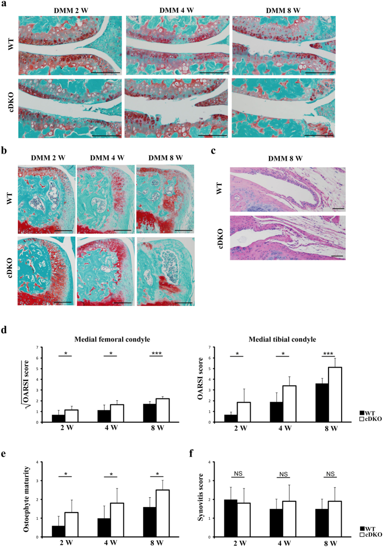
Osteoarthritis (OA) is a progressive degenerative disease of the joints that is associated with both joint injury and ageing. Here, we investigated the role of the energy sensor AMP-activated protein kinase (AMPK) in maintaining a healthy state of articular cartilage and in OA development. Using cartilage-specific, tamoxifen-inducible AMPKα1 conditional knockout (AMPKα1 cKO), AMPKα2 conditional knockout (AMPKα2 cKO) and AMPKα1α2 conditional double knockout (AMPKα cDKO) mice, we found that compared with wild-type (WT) littermates, mutant mice displayed accelerated severity of surgically induced OA, especially AMPKα cDKO mice. Furthermore, male but not female AMPKα cDKO mice exhibited severely spontaneous ageing-associated OA lesions at 12 months of age. The chondrocytes isolated from AMPKα cDKO mice resulted in an enhanced interleukin-1β (IL-1β)-stimulated catabolic response. In addition, upregulated expression of matrix metalloproteinase-3 (MMP-3), MMP-13 and phospho-nuclear factor-κB (phospho-NF-κB) p65 and increased levels of apoptotic markers were detected in the cartilage of AMPKα cDKO mice compared with their WT littermates in vivo. Thus, our findings suggest that AMPK activity in chondrocytes is important in maintaining joint homeostasis and OA development.


Conflict of interest statement

The authors declare no competing financial interests.

Figures

Figure 1
Figure 1. Efficiency of AMPK ablation in chondrocytes.
(a) Immunofluorescence analysis showed that AMPKα1 and AMPKα2 expressions levels were reduced in articular chondrocytes from 10-week-old AMPKα cDKO mice treated with TM at age 8 weeks for 5 days compared with the levels in their WT littermates (n = 6/group). Green indicates positive staining. Blue indicates DAPI staining. Scale bars = 50 μm. (b) Real-time reverse transcriptase-PCR analysis reveals significant reductions in AMPKα1 and AMPKα2 messenger RNA (mRNA) expression levels in cartilage obtained from 10-week-old AMPKα cDKO mice compared with the levels in their WT littermates (n = 6/group). These mice were treated with TM at age 8 weeks for 5 days. **p < 0.01.
Figure 2
Figure 2. Basal articular cartilage in AMPKα1α2 conditional double knockout (AMPKα cDKO) mice and their Cre-negative wild-type (WT) littermates.
(a) Safranin-O/Fast green and (b) H&E staining of knee joints from 10-week-old AMPKα cDKO mice and their WT littermates administrated TM at age 8 weeks for 5 days (n = 6/group). Scale bars = 100 μm. Representative IF images of (c) Col2a1 and (d) Sox9 in knee joints from 10-week-old AMPKα cDKO mice and their WT littermates administrated TM at age 8 weeks for 5 days (n = 6/group). Green indicates positive staining. Blue represents DAPI staining. No intensity or background adjustments were made between sections. Col2a1 = Type II collagen. Scale bars = 50 μm.
Figure 3
Figure 3. Accelerated OA in AMPKα1α2 conditional double knockout (AMPKα cDKO) mice following destabilization of the medial meniscus (DMM).
Representative photographs of articular cartilage destruction (a) or osteophyte formation (b) in AMPKα cDKO mice and their WT littermates 2, 4 and 8 weeks post-DMM (n = 10/group). Sections were stained with Safranin O/Fast Green. Scale bars = 100 μm. (c) Representative photographs of synovitis in AMPKα cDKO mice (n = 10) and their WT littermates (n = 10) at 8 weeks post-DMM. Sections were stained with H&E. Scale bars = 50 μm. The OARSI scores for the medial femoral condyle and the medial tibial condyle (d) and osteophyte maturity (e) at 2, 4 and 8 weeks post-DMM, and synovitis scores (f) at 8 weeks post-DMM in AMPKα cDKO mice and their WT littermates (n = 10/group). The OARSI scores for the medial femoral condyle of AMPKα cDKO and their WT littermates were transformed by taking the square root of the values. After transformation, all groups of data approximate a Gaussian distribution. *p < 0.05. **p < 0.01. ***p < 0.001. NS = not significant.
Figure 4
Figure 4. Accelerated OA in AMPKα1α2 conditional double knockout (AMPKα cDKO) mice with ageing.
(a) Representative photographs of articular cartilage destruction in female and male AMPKα cDKO mice and their age- and sex- matched WT littermates at 9 and 12 months of age. Sections were stained with Safranin O/Fast Green. A slight loss of proteoglycans in the superficial zone and fibrillation in articular cartilage (arrowhead) were observed in female AMPKα cDKO mice at 9 or 12 months. Loss of uncalcified cartilage and alteration of the tidemark integrity (arrow) were observed in male AMPKα cDKO mice at 9 months. Scale bars = 100 μm. (b) Representative Safranin-O/Fast green staining of knee joints from male AMPKα cDKO mice and their sex matched WT littermates at 12 months of age. Loss of the entire articular cartilage layer (black arrows), formation of osteophyte (green arrows), severe disruption of meniscal tissue (asterisk) and bone marrow-like regions in the enlarged medial collateral ligament (red arrows) were observed in male AMPKα cDKO mice at 12 months of age. T, tibia; F, femur; AC, articular cartilage; M, meniscus; MCL, medial collateral ligament. MAR, marrow cavities. Scale bars = 100 μm. (c) The OARSI scores for the medial femoral condyle and the medial tibial condyle in female and male AMPKα cDKO mice and their sex-matched WT littermates at 9 and 12 months of age (9 months old: WT females n = 5, cDKO females n = 7, WT males n = 7, cDKO males n = 9; 12 months old: WT females n = 7, cDKO females n = 8, WT males n = 8, cDKO males n = 11). (d) Representative photographs of synovitis in male AMPKα cDKO mice and their sex-matched WT littermates at 12 months of age. Sections were stained with H&E. Scale bars = 50 μm. (e) Osteophyte maturity and synovitis scores in male AMPKα cDKO mice (n = 11) and their sex-matched WT littermates (n = 8) at 12 months of age. *p < 0.05. **p < 0.01. ***p < 0.001. NS = not significant.
Figure 5
Figure 5. AMPKα deficiency enhancing phospho-NF-κB p65 and the procatabolic response to iterleukin-1β (IL-1β) in primary murine chondrocytes.
Primary articular chondrocytes were isolated from Col2a1-CreERT2; AMPKα1flox/floxα2flox/flox mice and their Cre-negative WT littermates and treated with 4-hydroxytamoxifen for 48 hours as described in the Methods. (a) Western blotting analyses of AMPKα expression. (b) The expression levels of Col2a1, Aggrecan, Sox9, MMP-3, MMP-13, Adamts4, Adamts5 and Timp3 messenger RNA (mRNA) in primary murine chondrocytes treated with or without IL-1β (10 ng/ml) for 24 h were determined by real-time reverse transcriptase-PCR. Data are representative of three individual experiments. Col2a1 = Type II collagen; MMP-3 = matrix metalloproteinase-3; MMP-13 = matrix metalloproteinase-13; *p < 0.05. ***p < 0.001. NS = not significant. (c) Western blotting analyses of total NF-κB p65 and phospho-NF-κB p65 in primary murine chondrocytes with or without IL-1β (10 ng/ml) for 24 h. Increased phospho-NF-κB p65 protein expression in chondrocytes from AMPKα cDKO mice compared with their WT littermates was observed. GAPDH served as a loading control.
Figure 6
Figure 6. IHC analyses of surgically induced and ageing-associated OA.
(a,c and e) Representative IHC images of MMP-3, MMP-13 and phospho-NF-κB p65 in the medial tibial plateau in AMPKα1α2 conditional double knockout (AMPKα cDKO) mice and their WT littermates 2 weeks post-sham operation and DMM surgery or in mice at 9 months of age. Scale bars = 20 μm. The cellularity of the section was confirmed with haematoxylin staining. (b,d,f) Quantifications of the percentages of MMP-3, MMP-13 and phospho-NF-κB p65 are presented as percentages relative to cells stained for haematoxylin. (n = 6/group) MMP-3 = matrix metalloproteinase-3; MMP-13 = matrix metalloproteinase-13. *p < 0.05. **p < 0.01. ***p < 0.001. NS = not significant.
Figure 7
Figure 7. TUNEL analyses of surgically induced and ageing-associated OA.
(a) Representative TUNEL images in the medial tibial plateau in AMPKα1α2 conditional double knockout (AMPKα cDKO) mice and their WT littermates 2 weeks post-sham operation and DMM surgery or in mice at 9 months of age. Green represents positive staining. Blue indicates DAPI staining. No intensity or background adjustments were made between sections. Scale bars = 50 μm. (b) Quantifications of the percentage of TUNEL-positive cells are presented as percentages relative to cells stained for DAPI. (n = 6/group) TUNEL = terminal deoxynucleotidyl transferase dUTP nick end labelling; **p < 0.01. ***p < 0.001. NS = not significant.

Similar articles

Cited by

References

    1. Aigner T., Sachse A., Gebhard P. M. & Roach H. I. Osteoarthritis: pathobiology-targets and ways for therapeutic intervention. Adv. Drug Deliv. Re. 58, 128–149 (2006). - PubMed
    1. Loeser R. F., Goldring S. R., Scanzello C. R. & Goldring M. B. Osteoarthritis: a disease of the joint as an organ. Arthritis Rheum. 64, 1697–1707 (2012). - PMC - PubMed
    1. Witczak C. A., Sharoff C. G. & Goodyear L. J. AMP-activated protein kinase in skeletal muscle: from structure and localization to its role as a master regulator of cellular metabolism. Cell Mol. Life. Sci. 65, 3737–3755 (2008). - PMC - PubMed
    1. Steinberg G. R. & Kemp B. E. AMPK in Health and Disease. Physiol. Rev. 89, 1025–1078 (2009). - PubMed
    1. Salminen A. & Kaarniranta K. AMP-activated protein kinase (AMPK) controls the aging process via an integrated signaling network. Ageing Res. Rev. 11, 230–241 (2012). - PubMed

Publication types

Substances