

Spherical Soft Contact Lens Designs and Peripheral Defocus in Myopic Eyes
- PMID: 28225372
- PMCID: PMC5324711
- DOI: 10.1097/OPX.0000000000001053
Spherical Soft Contact Lens Designs and Peripheral Defocus in Myopic Eyes
Abstract
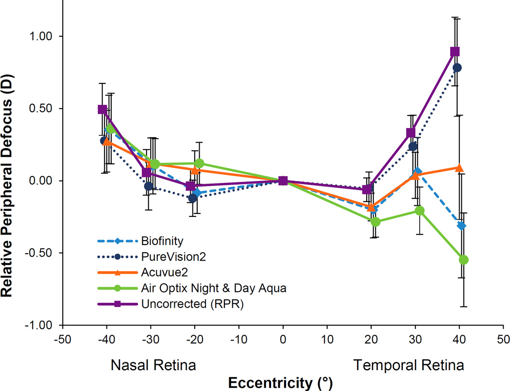
Purpose: Peripheral retinal defocus has been implicated in myopia progression. The effect of commercially available spherical soft contact lenses (SCLs) on peripheral defocus of adult myopic eyes was investigated.
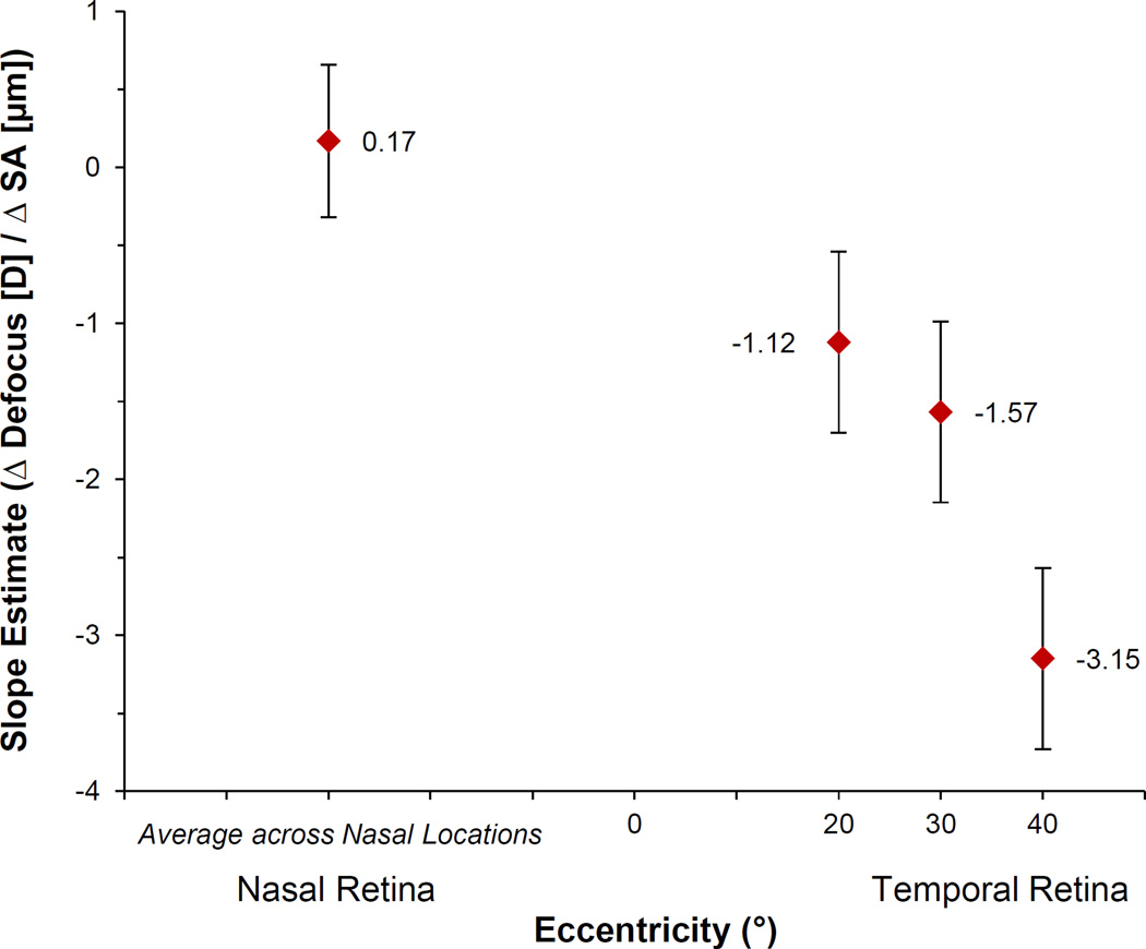
Methods: Twenty-five young adults with spherical equivalent (SE) refractions between -0.50D and -6.00D were enrolled. Cycloplegic autorefraction (right eye) was measured centrally and ±20°, ±30°, and ±40° from the line of sight along the horizontal meridian using an autorefractor. Four commercially available spherical SCLs (Biofinity, Acuvue2, PureVision2, and Air Optix Night & Day Aqua) were evaluated. SE defocus (M) was used to calculate relative peripheral defocus (RPD) while wearing each SCL and relative peripheral refraction of the uncorrected eye. Spherical aberration (SA) changes caused by each SCL were measured along the line of sight by aberrometry. Peripheral defocus was analyzed using repeated-measures analyses of variance (RM-ANOVA). The association between changes in axial SA and the change in peripheral defocus was evaluated using linear mixed models.
Results: The mean age (±SD) and central SE refractive error were 24.0 ± 1.3 years and -3.45 ± 1.42D, respectively. PureVision2 did not change RPD (P = .33). Significant myopic shifts on the temporal retina were found with three lenses: Acuvue 2 (-0.29D at 30°; -0.80D at 40°; both P ≤ .01), Biofinity (-1.21 D at 40°; P = .02), and Air Optix Night & Day Aqua (-0.23D at 20°, -0.48D at 30°, and -1.50D at 40°; all P < .004). All SCLs caused a negative change in SA. SCLs inducing less negative (more positive) SA changes were associated with a less hyperopic change in RPD.
Conclusions: Spherical SCL design can influence the peripheral defocus profile experienced by a myopic eye. Several, but not all, SCLs reduced peripheral hyperopia. Differences in how SCL types influence peripheral defocus may have implications for myopia progression.
Figures
References
-
- Lin LL, Shih YF, Tsai CB, et al. Epidemiologic study of ocular refraction among schoolchildren in Taiwan in 1995. Optom Vis Sci. 1999;76:275–281. - PubMed
-
- Morgan IG, Ohno-Matsui K, Saw SM. Myopia. Lancet. 2012;379:1739–1748. - PubMed
-
- Holden BA, Fricke TR, Wilson DA, et al. Global prevalence of myopia and high myopia and temporal trends from 2000 through 2050. Ophthalmology. 2016;123:1036–1042. - PubMed
-
- Saw SM, Gazzard G, Shih-Yen EC, et al. Myopia and associated pathological complications. Ophthalmic Physiol Opt. 2005;25:381–391. - PubMed
MeSH terms
Grants and funding
LinkOut - more resources
Full Text Sources
Other Literature Sources
Research Materials
Miscellaneous
