. 2017 Apr;158(4):747-759.
doi: 10.1097/j.pain.0000000000000830.

Divergent functions of the left and right central amygdala in visceral nociception

Affiliations

Divergent functions of the left and right central amygdala in visceral nociception

Katelyn E Sadler et al. Pain. 2017 Apr.

Abstract

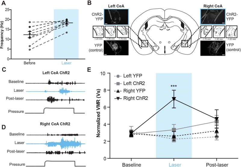
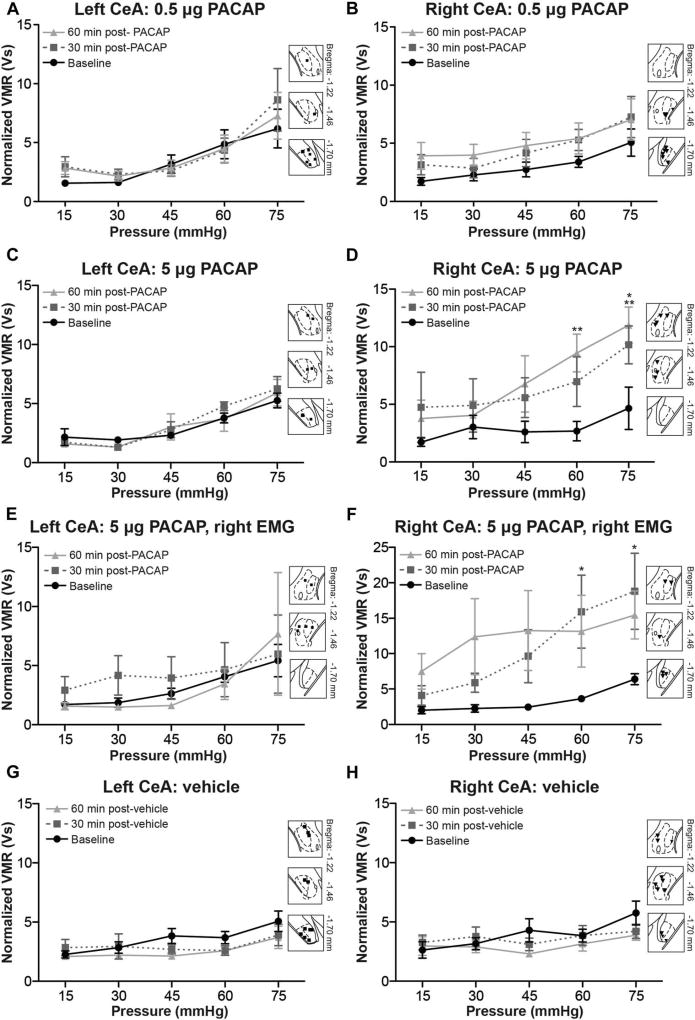
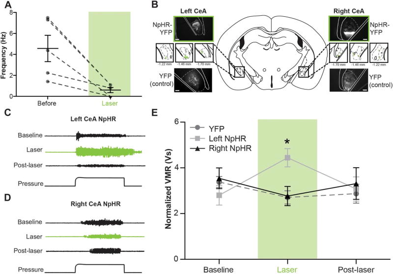
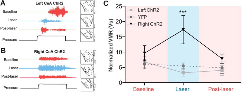
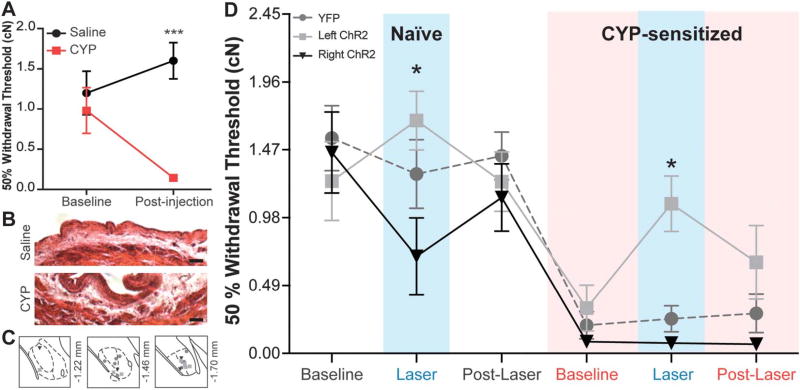
The left and right central amygdalae (CeA) are limbic regions involved in somatic and visceral pain processing. These 2 nuclei are asymmetrically involved in somatic pain modulation; pain-like responses on both sides of the body are preferentially driven by the right CeA, and in a reciprocal fashion, nociceptive somatic stimuli on both sides of the body predominantly alter molecular and physiological activities in the right CeA. Unknown, however, is whether this lateralization also exists in visceral pain processing and furthermore what function the left CeA has in modulating nociceptive information. Using urinary bladder distension (UBD) and excitatory optogenetics, a pronociceptive function of the right CeA was demonstrated in mice. Channelrhodopsin-2-mediated activation of the right CeA increased visceromotor responses (VMRs), while activation of the left CeA had no effect. Similarly, UBD-evoked VMRs increased after unilateral infusion of pituitary adenylate cyclase-activating polypeptide in the right CeA. To determine intrinsic left CeA involvement in bladder pain modulation, this region was optogenetically silenced during noxious UBD. Halorhodopsin (NpHR)-mediated inhibition of the left CeA increased VMRs, suggesting an ongoing antinociceptive function for this region. Finally, divergent left and right CeA functions were evaluated during abdominal mechanosensory testing. In naive animals, channelrhodopsin-2-mediated activation of the right CeA induced mechanical allodynia, and after cyclophosphamide-induced bladder sensitization, activation of the left CeA reversed referred bladder pain-like behaviors. Overall, these data provide evidence for functional brain lateralization in the absence of peripheral anatomical asymmetries.


Conflict of interest statement

Conflict of interest statement

The authors have no conflicts of interest to declare.

Figures

Figure 1
Figure 1. Optogenetic activation of the right central amygdala (CeA) increases pain-like responses to noxious bladder distension
(A) Pulsing 473-nm light at 20 Hz significantly increased firing rates of neurons in channelrhodopsin-2 (ChR2)–expressing regions of the CeA (paired t test, P = 0.0009; n = 9). (B) Three weeks after targeted viral injections, nestin-Cre mice expressed either yellow fluorescent protein (YFP)–tagged ChR2 or YFP alone in the left or right CeA (scale bar, 0.1 mm). Targeting for all experimental and control animals is shown in insets (blue ■, left ChR2-YFP; gray ●, left YFP; blue ▼, right ChR2-YFP; gray ▲, right YFP). (C) Representative traces show electromyographic responses to noxious bladder distension (60 mm Hg) prior to, during, and after laser-induced activation of ChR2-expressing neurons in the left CeA. Optogenetic activation failed to produce significant qualitative changes in abdominal muscle output. (D) Optogenetic activation of the right CeA induced marked increases in abdominal muscle contraction during bladder distension. (E) Optogenetic activation of the right CeA significantly increased visceromotor responses (VMRs) (2-way repeated measures analysis of variance effect of group, P = 0.0296; effect of time, P = 0.0330; group × time interaction, P = 0.0001; Bonferroni posttest right ChR2 vs right YFP at laser, ***P < 0.001; right ChR2 laser vs baseline, ***P < 0.001; right ChR2 vs left ChR2 at laser, ***P < 0.001; n = 6 in each group). Optogenetic activation of the left CeA had no significant effect on VMRs during bladder distension, and likewise, laser stimulation in control groups had no effect on VMRs. Fifteen minutes after the end of laser stimulation, VMRs for all experimental and control groups were similar to each other and not significantly different from baseline responses.
Figure 2
Figure 2. Left and right central amygdalae (CeA) do not differ in neuron number or activation levels during channelrhodopsin-2 (ChR2)–mediated depolarization
(A) NeuN staining was used to determine the number of neurons in the left and right CeA of naive C57Bl/6 animals. The NeuN signal does not differ between nuclei (paired t test, P = 0.8238; n = 8); matching colored symbols represent the average left and right signal from one animal. Insets show representative images of each nucleus. (B) The left and right CeA of animals expressing ChR2-yellow fluorescent protein (YFP) were stained for cFos after optogenetic stimulation and urinary bladder distension. cFos-positive cells are predominantly located in the CeA (scale bar, 0.1 mm). (C) cFos was also examined in both the injected and noninjected sides of brains from animals expressing YFP alone (scale bar, 0.1 mm). (D) Both left ChR2 (n = 11) and right ChR2 (n = 7)-expressing nuclei exhibited increases in cFos expression when compared with their nonstimulated counternuclei (2-way repeated measures analysis of variance effect of injection, P < 0.0001; group × injection interaction, P = 0.0144; Bonferroni posttest for left ChR2 injected vs noninjected, ***P < 0.001; right ChR2 injected vs noninjected, **P < 0.01). Additionally, the activation levels between these left and right stimulated nuclei do not differ. In YFP-expressing brains (n = 6), there is no significant difference in cFos expression between injected and noninjected nuclei.
Figure 3
Figure 3. Pituitary adenylate cyclase–activating polypeptide (PACAP) administration in the right central amygdala (CeA), and not the left, increases responses to bladder distension. Two doses of PACAP were administered into the left or right CeA after one round of graded urinary bladder distension (UBD)
(A) Infusion of 0.5 µg PACAP into the left CeA failed to change visceromotor responses (VMRs) 30 or 60 minutes after application (2-way repeated measures [RM] analysis of variance [ANOVA] effect of pressure, P = 0.0041; n = 9) Insets show targeting of the left CeA. (B) Infusion of 0.5 µg PACAP in the right CeA significantly increased VMRs (2-way RM ANOVA effect of treatment, P = 0.0021; effect of pressure, P = 0.0220; n=9). Insets show targeting of the right CeA. (C) Infusion of 5 µg PACAP dose in the left CeA failed to significantly change VMRs obtained from the left abdominal muscle 30 or 60 minutes after application (2-way RM ANOVA effect of pressure, P < 0.0001; n = 6). (D) Infusion of 5 µg PACAP into the right CeA resulted in statistically significant increases in UBD VMRs recorded from the left abdominal muscle, specifically at the most noxious intravesical pressures (2-way RM ANOVA effect of treatment, P < 0.0001; effect of pressure, P = 0.0767; Bonferroni posttest for baseline vs 30 minutes, *P <0.05; for baseline vs 60 minutes, **P < 0.01; n = 6). (E) VMRs from the right abdominal muscle did not change after administration of 5 µg PACAP in the left CeA. (F) Infusion of 5 µg PACAP in the right CeA causes a robust increase in UBD-evoked VMRs recorded from the right abdominal muscle (2-way RM ANOVA effect of treatment, P < 0.0001; n = 6). Significant increases in UBD-evoked VMRs were noted 30 minutes after PACAP administration (Bonferroni posttest for baseline vs 30 minutes, *P < 0.05 at 60 and 75 mm Hg). (G, H) To control for microinjection manipulation and time elapsed during procedure, vehicle (0.05% bovine serum albumin in saline) was applied into the left or right CeA in a third group of animals. No change in VMR occurred 30 or 60 minutes after vehicle application in either (G) the left CeA (2-way RM ANOVA effect of pressure, P = 0.0001; n = 11) or (H) the right CeA (2-way RM ANOVA effect of pressure, P = 0.0034; n = 10).
Figure 4
Figure 4. Graded bladder distension equally activates left and right para-brachial nucleus (PBn)
(A) cFos-positive neurons were localized to the left and right lateral PBn after one round of graded bladder distension. (B) Urinary bladder distension (UBD) significantly and equivalently increased the number of cFos-positive cells in the left and right PBn compared with sham animals (2-way repeated measures analysis of variance effect of UBD, P = 0.0018; n = 6; scale bar, 0.05 mm).
Figure 5
Figure 5. Optogenetic inhibition of the left central amygdala (CeA) increases pain-like responses to noxious bladder distension
(A) Constant application of 532-nm light in NpHR-expressing regions of the CeA significantly decreased neuronal firing rates (paired t test, P = 0.0224; n = 5). (B) Three weeks after targeted viral injections, nestin-Cre mice expressed either yellow fluorescent protein (YFP)–tagged NpHR or YFP alone in the left or right CeA (scale bar, 0.1 mm). Targeting for all experimental and control animals is shown in insets (green ■, left NpHR-YFP; gray ●, left YFP; green ▼, right NpHR-YFP; gray ▲, right YFP). (C) Representative electromyographic (EMG) traces show qualitative changes during left CeA optogenetic inhibition. (D) Representative EMG traces fail to show qualitative changes during right CeA optogenetic inhibition. (E) Quantified visceromotor responses (VMRs) show that optogenetic inhibition of the left CeA significantly increased responses to urinary bladder distension when compared with VMRs obtained during right CeA inhibition or optical stimulation in YFP-expressing CeA (2-way repeated measures analysis of variance group × time interaction, P = 0.0126; Bonferroni posttest left NpHR vs right NpHR at laser, *P < 0.05, left NpHR vs YFP at laser, *P < 0.05, left NpHR laser vs baseline, *P < 0.05; n = 9–19). Fifteen minutes after the end of laser stimulation, VMRs were once again equivalent between groups and not different from baseline responses.
Figure 6
Figure 6. Optogenetic activation of the right central amygdala (CeA) in bladder-sensitized mice further increases bladder nociception. After cyclophosphamide treatment, animals expressing channelrhodopsin-2 (ChR2) in the left or right CeA were placed through 3 rounds of noxious (60 mm Hg) bladder distension
(A) Representative electromyographic (EMG) traces during optogenetic activation of the left CeA. Insets illustrate viral targeting in the CeA (gray ■, left ChR2). (B) Representative EMG traces during right CeA optogenetic activation display increases in visceromotor responses (VMRs). Insets illustrate viral targeting in the CeA (black ▼, right ChR2). (C) In bladder-sensitized animals, optogenetic activation of the right CeA significantly increased urinary bladder distension–evoked VMRs (2-way repeated measures analysis of variance overall effect of group, P = 0.0218; group × time interaction, P = 0.0264; Bonferroni posttest for right ChR2 vs yellow fluorescent protein (YFP) at laser, **P < 0.01; right ChR2 vs left ChR2 at laser, ***P < 0.001; right ChR2 baseline vs laser, *P < 0.05; n = 5–7). Laser stimulation in YFP-expressing control mice had no effect on VMRs.
Figure 7
Figure 7. Activation of the right central amygdala (CeA) increases naive abdominal sensitivity while activation of the left CeA reverses referred bladder pain. Five days of cyclophosphamide (CYP) treatment induces referred bladder pain as measured by von Frey filaments
(A) CYP-treated animals display a statistically significant decrease in abdominal withdrawal threshold when compared with saline-treated animals (2-way repeated measures [RM] analysis of variance [ANOVA] overall effect of CYP, P = 0.0042; effect of interaction, P = 0.0030; Bonferroni posttest CYP vs saline at postinjection, ***P < 0.001; n = 12). (B) CYP-treated animals display noticeable edema in the submucosal layers of the bladder and thickening of the urothelium (scale bar, 50 µm). (C) channelrhodopsin-2 (ChR2) and yellow fluorescent protein (YFP) vectors were successfully targeted to the left or right CeA (gray ●, YFP; light gray ■, left ChR2; black ▼, right ChR2). (D) In naive animals, optogenetic activation of the right CeA causes a significant decrease in withdrawal threshold compared with the effects of left CeA activation (n = 6–9; 2-way ANOVA effect of group, P = 0.0017; Bonferroni posttest right ChR2 vs left ChR2 at naive laser, *P < 0.05), suggesting divergent effects of the 2 nuclei. Withdrawal thresholds of each group do not differ prior to and after laser-induced activation of either nucleus. After 5 days of CYP treatment, all groups exhibit referred bladder pain as indicated by significantly reduced mechanical withdrawal thresholds (2-way RM ANOVA effect of time, P < 0.0001; Bonferroni posttest naive baseline vs CYP baseline for YFP, P < 0.0001; for left ChR2, P < 0.05; for right ChR2, P < 0.01). Optogenetic activation of the right CeA has no assessable effect on withdrawal thresholds in sensitized animals. Optogenetic activation of the left CeA during CYP sensitization reverses referred bladder pain (Bonferroni posttest left ChR2 vs right ChR2 at CYP laser, *P < 0.05).

References

    1. As-Sanie S, Harris RE, Napadow V, Kim J, Neshewat G, Kairys A, Williams D, Clauw DJ, Schmidt-Wilcke T. Changes in regional gray matter volume in women with chronic pelvic pain: a voxel-based morphometry study. PAIN. 2012;153:1006–14. - PMC - PubMed
    1. Baas D, Aleman A, Kahn RS. Lateralization of amygdala activation: a systematic review of functional neuroimaging studies. Brain Res Brain Res Rev. 2004;45:96–103. - PubMed
    1. Bagarinao E, Johnson KA, Martucci KT, Ichesco E, Farmer MA, Labus J, Ness TJ, Harris R, Deutsch G, Apkarian AV, Mayer EA, Clauw DJ, Mackey S. Preliminary structural MRI based brain classification of chronic pelvic pain: a MAPP network study. PAIN. 2014;155:2502–9. - PMC - PubMed
    1. Bauer RH. Lateralization of neural control for vocalization by the frog (Rana pipiens) Psychobiology. 1993;21:243–8.
    1. Bon K, Lanteri-Minet M, Michiels JF, Menetrey D. Cyclophosphamide cystitis as a model of visceral pain in rats: a c-fos and Krox-24 study at telencephalic levels, with a note on pituitary adenylate cyclase activating polypeptide (PACAP) Exp Brain Res. 1998;122:165–74. - PubMed

Publication types

MeSH terms