. 2017 Feb 22;12(2):e0172477.
doi: 10.1371/journal.pone.0172477. eCollection 2017.

Capsaicin reduces Alzheimer-associated tau changes in the hippocampus of type 2 diabetes rats

Affiliations

Capsaicin reduces Alzheimer-associated tau changes in the hippocampus of type 2 diabetes rats

Weijie Xu et al. PLoS One. .

Abstract

Type 2 diabetes (T2D) is a high-risk factor for Alzheimer's disease (AD) due to impaired insulin signaling pathway in brain. Capsaicin is a specific transient receptor potential vanilloid 1 (TRPV1) agonist which was proved to ameliorate insulin resistance. In this study, we investigated whether dietary capsaicin could reduce the risk of AD in T2D. T2D rats were fed with capsaicin-containing high fat (HF) diet for 10 consecutive days (T2D+CAP). Pair-fed T2D rats (T2D+PF) fed with the HF-diet of average dose of T2D+CAP group were included to control for the effects of reduced food intake and body weight. Capsaicin-containing standard chow was also introduced to non-diabetic rats (NC+CAP). Blood glucose and insulin were monitored. The phosphorylation level of tau at individual sites, the activities of phosphatidylinositol 3 kinase/protein kinase B (PI3K/AKT) and glycogen synthase kinase-3β (GSK-3β) were analyzed by Western blots. The results revealed that the levels of phosphorylated tau protein at sites Ser199, Ser202 and Ser396 in hippocampus of T2D+CAP group were decreased significantly, but these phospho-sites in T2D+PF group didn't show such improvements compared with T2D group. There were almost no changes in non-diabetic rats on capsaicin diet (NC+CAP) compared with the non-diabetic rats with normal chow (NC). Increased activity of PI3K/AKT and decreased activity of GSK-3β were detected in hippocampus of T2D+CAP group compared with T2D group, and these changes did not show in T2D+PF group either. These results demonstrated that dietary capsaicin appears to prevent the hyperphosphorylation of AD-associated tau protein by increasing the activity of PI3K/AKT and inhibiting GSK-3β in hippocampus of T2D rats, which supported that dietary capsaicin might have a potential use for the prevention of AD in T2D.


Conflict of interest statement

Competing Interests: The funder Novo Nordisk had no role in study design, data collection and analysis, decision to publish, or preparation of the manuscript. The authors declare that there is no financial or non-financial competing interests with the founder Novo Nordisk. This does not alter our adherence to PLOS ONE policies on sharing data and materials.

Figures

Fig 1
Fig 1. The diagram for experimental design.
60 male SD rats were included in this study. 40 rats were assigned to T2D model group and fed by HF diet, while 20 rats were assigned to control group and fed by standard chow. 12 weeks later, rats in T2D model group were injected with STZ, while rats in the control group were injected with citrate buffer only. The whole blood glucose and plasma insulin were monitored 3 days after the STZ or citrate buffer injection. Hyperglycemia was verified 3 days after the STZ injection, 8 rats did not develop diabetes within 3 days after the STZ injection were excluded from further assessment. Thereafter, capsaicin-containing HF diet was introduced to T2D+CAP group (n = 11), rats in T2D+PF (n = 11) group were pair-fed with HF diet according to the amount of HF diet intake of T2D+CAP group one day before, and T2D group (n = 10) got free access to HF diet. For control group, half of the rats were introduced to capsaicin-containing standard chow (NC+CAP, n = 10) while another half (NC, n = 10) still got free access to standard chow. After 10 days of capsaicin exposure, whole blood glucose and plasma insulin were monitored again before sacrifice.
Fig 2
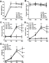
Fig 2. Changes of body weight (2A), energy intake (2B), blood glucose (2C), plasma insulin (2D) and HOMA-IR (2E).
Data are mean ± SD. Differences vs. NC group are indicated as *P < 0.05, **P < 0.01; differences vs. T2D group are indicated as ⊿P < 0.05 and differences vs. T2D+PF group are indicated as #P < 0.05. ‘NC’ indicated non-diabetic rats fed with standard chow, ‘NC+CAP’ indicated non-diabetic rats fed with capsaicin-containing standard chow, ‘T2D’ indicated T2D rats with free access to HF diet, ‘T2D+PF’ indicated T2D rats pair-fed with HF diet according to the amount of HF diet intake of T2D+CAP group one day before. ‘T2D+CAP’ indicated T2D rats fed with capsaicin-containing HF diet. Measurements were taken on week -12, day -3, day 0 and day 10.
Fig 3
Fig 3. Western blotting analysis of phosphorylation of tau protein in rat hippocampus.
3A, western blots of the crude hippocampal extracts (7–13 μg/lane) were analyzed with several site-specific tau antibodies to detect the phosphorylation levels of tau at the specific sites. Each lane was from an individual rat. Actin blot was included as a loading control. 3B, the blots as shown in panel 3A were quantitated desitometrically and for quantitation of tau phosphorylation level at each site, data had been normalized by the level of total tau. All data are presented as mean ± SD of the relative immunoreactivities. *P < 0.05,**P < 0.01 as compared to NC group; ⊿P < 0.05 as compared to T2D group and #P < 0.05 as compared to T2D+PF group.
Fig 4
Fig 4. Western blot analysis of insulin signal transduction in rat hippocampus.
Crude hippocampal extracts (5–15 μg/lane) were analyzed by Western blots developed with antibodies to total AKT and phosph-AKT (4A, B), total GSK3β and phosph-GSK3β (4C, D). Actin blot was included as a loading control. Each lane was from an individual rat (4A, C), the blots as shown in panel 4B, D were quantitated desitometrically and for quantitation of total AKT and total GSK-3β. All data are presented as mean ± SD of the relative immunoreactivities. *P < 0.05,**P < 0.01 as compared to NC group; ⊿P < 0.05 as compared to T2D group and #P < 0.05 as compared to T2D+PF group.

References

    1. Henry MS, Passmore AP, Todd S, McGuinness B, Craig D, Johnston JA. The development of effective biomarkers for Alzheimer's disease: a review. Int J Geriatr Psychiatry. 2013;28(4):331–40. Epub 2012/06/08. 10.1002/gps.3829 - DOI - PubMed
    1. Grundke-Iqbal I, Iqbal K, Tung YC, Quinlan M, Wisniewski HM, Binder LI. Abnormal phosphorylation of the microtubule-associated protein tau (tau) in Alzheimer cytoskeletal pathology. Proc Natl Acad Sci U S A. 1986;83(13):4913–7. Epub 1986/07/01. - PMC - PubMed
    1. Grundke-Iqbal I, Iqbal K, Quinlan M, Tung YC, Zaidi MS, Wisniewski HM. Microtubule-associated protein tau. A component of Alzheimer paired helical filaments. J Biol Chem. 1986;261(13):6084–9. Epub 1986/05/05. - PubMed
    1. Arriagada PV, Growdon JH, Hedley-Whyte ET, Hyman BT. Neurofibrillary tangles but not senile plaques parallel duration and severity of Alzheimer's disease. Neurology. 1992;42(3 Pt 1):631–9. Epub 1992/03/01. - PubMed
    1. Gomez-Isla T, Hollister R, West H, Mui S, Growdon JH, Petersen RC, et al. Neuronal loss correlates with but exceeds neurofibrillary tangles in Alzheimer's disease. Ann Neurol. 1997;41(1):17–24. Epub 1997/01/01. 10.1002/ana.410410106 - DOI - PubMed

MeSH terms