

Ex vivo activation of CD4+ T-cells from donors on suppressive ART can lead to sustained production of infectious HIV-1 from a subset of infected cells
- PMID: 28225830
- PMCID: PMC5338860
- DOI: 10.1371/journal.ppat.1006230
Ex vivo activation of CD4+ T-cells from donors on suppressive ART can lead to sustained production of infectious HIV-1 from a subset of infected cells
Abstract
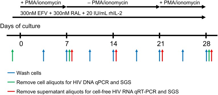
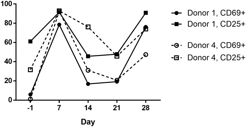
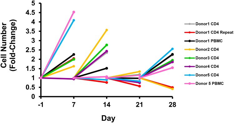
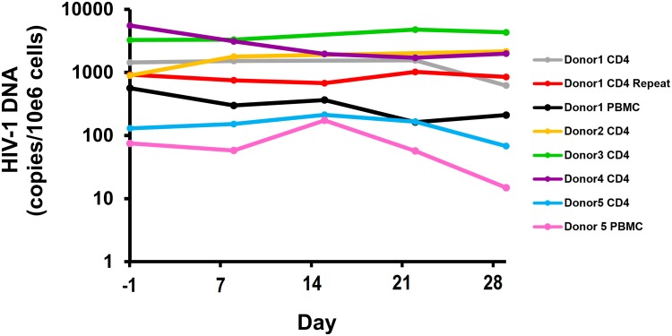
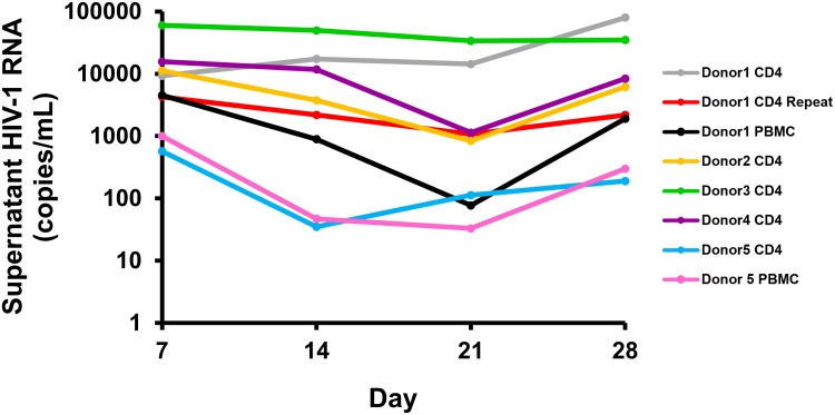
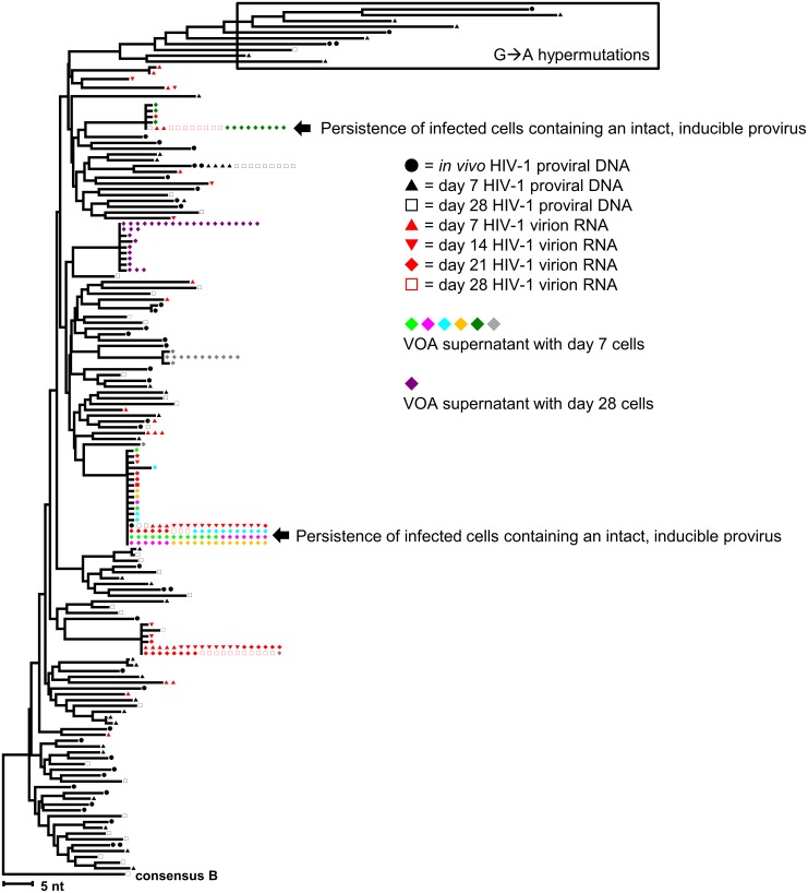
The fate of HIV-infected cells after reversal of proviral latency is not well characterized. Simonetti, et al. recently showed that CD4+ T-cells containing intact proviruses can clonally expand in vivo and produce low-level infectious viremia. We hypothesized that reversal of HIV latency by activation of CD4+ T-cells can lead to the expansion of a subset of virus-producing cells rather than their elimination. We established an ex vivo cell culture system involving stimulation of CD4+ T-cells from donors on suppressive antiretroviral therapy (ART) with PMA/ionomycin (day 1-7), followed by rest (day 7-21), and then repeat stimulation (day 21-28), always in the presence of high concentrations of raltegravir and efavirenz to effectively block new cycles of viral replication. HIV DNA and virion RNA in the supernatant were quantified by qPCR. Single genome sequencing (SGS) of p6-PR-RT was performed to genetically characterize proviruses and virion-associated genomic RNA. The replication-competence of the virions produced was determined by the viral outgrowth assay (VOA) and SGS of co-culture supernatants from multiple time points. Experiments were performed with purified CD4+ T-cells from five consecutively recruited donors who had been on suppressive ART for > 2 years. In all experiments, HIV RNA levels in supernatant increased following initial stimulation, decreased or remained stable during the rest period, and increased again with repeat stimulation. HIV DNA levels did not show a consistent pattern of change. SGS of proviruses revealed diverse outcomes of infected cell populations, ranging from their apparent elimination to persistence and expansion. Importantly, a subset of infected cells expanded and produced infectious virus continuously after stimulation. These findings underscore the complexity of eliminating reservoirs of HIV-infected cells and highlight the need for new strategies to kill HIV-infected cells before they can proliferate.
Conflict of interest statement
I have read the journal's policy and the authors of this manuscript have the following competing interests: JWM is a consultant for Gilead Sciences and a shareholder of Cocrystal Pharma, Inc. WS and BL are employees of Leidos Biomedical Research, Inc.
Figures
References
-
- Finzi D, Hermankova M, Pierson T, Carruth LM, Buck C, Chaisson RE, et al. Identification of a reservoir for HIV-1 in patients on highly active antiretroviral therapy. Science. 1997;278(5341):1295–300. - PubMed
-
- Wong JK, Hezareh M, Gunthard HF, Havlir DV, Ignacio CC, Spina CA, et al. Recovery of replication-competent HIV despite prolonged suppression of plasma viremia. Science. 1997;278(5341):1291–5. - PubMed
Publication types
MeSH terms
Substances
Grants and funding
LinkOut - more resources
Full Text Sources
Other Literature Sources
Medical
Research Materials
