

Review
doi: 10.1590/abd1806-4841.20175514.
Diabetes mellitus and the skin
Affiliations
- PMID: 28225950
- PMCID: PMC5312172
- DOI: 10.1590/abd1806-4841.20175514
Item in Clipboard
Review
Diabetes mellitus and the skin
An Bras Dermatol.
2017 Jan-Feb.
Abstract
Several dermatoses are routinely associated with diabetes mellitus, especially in patients with chronic disease. This relationship can be easily proven in some skin disorders, but it is not so clear in others. Dermatoses such necrobiosis lipoidica, granuloma annulare, acanthosis nigricans and others are discussed in this text, with an emphasis on proven link with the diabetes or not, disease identification and treatment strategy used to control those dermatoses and diabetes.
Conflict of interest statement
Conflict of interest: none
Figures
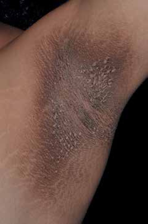
Armpits are a classic location of acanthosis nigricans. Note the
thickening and hyperchromia of the skin Photo: Department of Dermatology, Botucatu Medical School,
UNESP
Bullosis diabeticorum blisters are asymptomatic and exhibit mild
inflammation Photo: Department of Dermatology, Botucatu Medical School,
UNESP
Diabetic dermopathy consists of small brownish-colored depressions in the
skin surface, of atrophic appearance, which look like scars Photo: Department of Dermatology, Botucatu Medical School,
UNESP
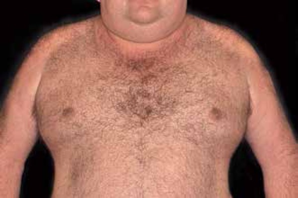
Buschke's scleredema is characterized by symmetrical anddiffuse
thickening, with hardening of the skin mainly on the face, cervical
region and upper limbs Photo: Department of Dermatology, Botucatu Medical School,
UNESP
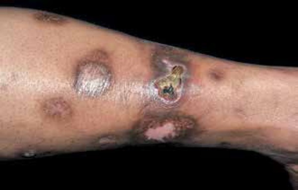
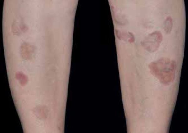
Granuloma annulare manifests by erythematous and firm dermal papules that
expand gradually, with central hyperpigmentation Photo: Department of Dermatology, Botucatu Medical School,
UNESP
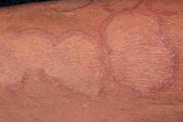
Detail of the granuloma annulare, showing infiltration at the edges of
the lesion Photo: Department of Dermatology, Botucatu Medical School,
UNESP
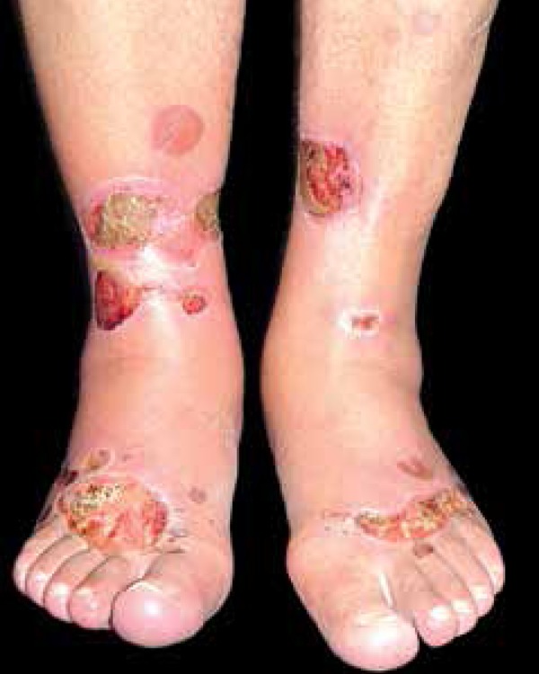
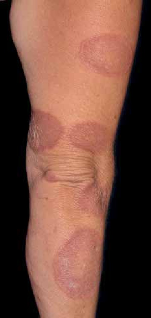
Typical lesions of necrobiosis lipoidica begin in the pretibial regions
with non-squamous papules that gradually grow and group into large
plaques Photo: Department of Dermatology, Botucatu Medical School,
UNESP
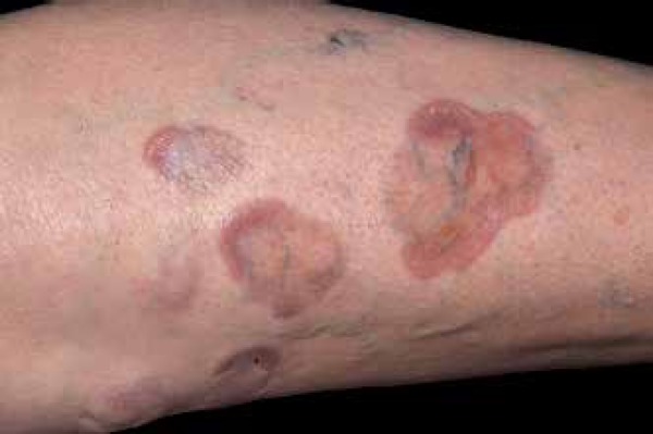
Detail of lesions of necrobiosis lipoidica, showing central atrophy Photo: Department of Dermatology, Botucatu Medical School,
UNESP
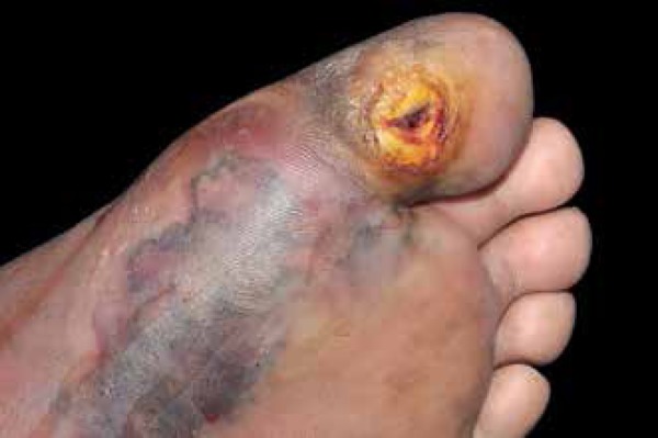
Diabetic foot may present a chronic ulcer on callus caused by changes in
sensitivity associated with diabetic neuropathy and occasional
ischemia Photo: Department of Dermatology, Botucatu Medical School,
UNESP
References
-
- Idf.org IDF Diabetes Atlas. 6th ed. [2015 Jul 15]. Internet. Available from: http://www.idf.org/diabetesatlas/update2014.
-
- Callen JP, Jorizzo JL, Bolognia JL, Piette WW, Zone JJ. Dermatological Signs of Internal Disease. 4th ed. Philadelphia: Saunders Elsevier; 2009.
-
- Barbato MT, Criado PR, Silva AK, Averbeck E, Guerine MB, Sá NB. Association of acanthosis nigricans and skin tags with insulin resistance. An Bras Dermatol. 2012;87:97–104. - PubMed
-
- Tamega Ade A, Aranha AM, Guiotoku MM, Miot LD, Miot HA. Association between skin tags and insulin resistance. An Bras Dermatol. 2010;85:25–31. - PubMed
-
- Papa CM. Niacinamide and acanthosis nigricans. Arch Dermatol. 1984;120:1281–1281. - PubMed
Publication types
MeSH terms
LinkOut - more resources
Full Text Sources
Other Literature Sources
Medical