

Case Reports
doi: 10.1590/abd1806-4841.20173563.
Hemosiderotic dermatofibroma
Affiliations
- PMID: 28225963
- PMCID: PMC5312185
- DOI: 10.1590/abd1806-4841.20173563
Item in Clipboard
Case Reports
Hemosiderotic dermatofibroma
An Bras Dermatol.
2017 Jan-Feb.
Abstract
We report a rare clinical case of hemosiderotic dermatofibroma in a 36-year-old female patient. The main dermatoscopic finding was represented by homogeneous blue-gray pigmentation. The aim of this report is to demonstrate the rarity of the lesion and the dermatoscopic importance it assumes by sharing a blue-gray homogeneous pattern with other benign and malignant lesions.
Conflict of interest statement
Conflict of Interest: None
Figures
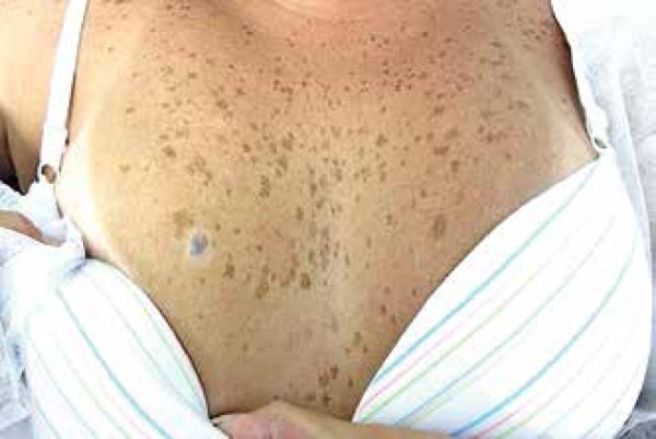
Blue-gray hard plaque with a hypochromic halo on the right breast
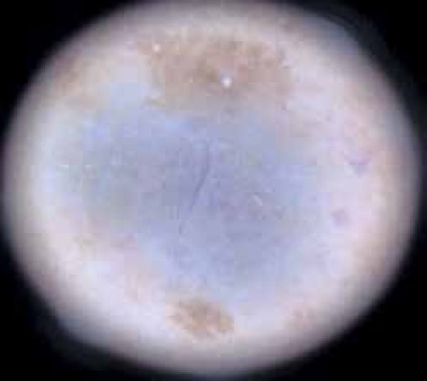
Dermatoscopic examination revealed a homogeneous blue-gray area and
collision of freckles at the periphery of the lesion
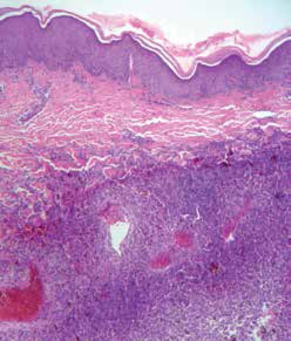
Histopathology: dermal involvement represented by the proliferation of
cells, some showing brown pigmentation and other involving thickened
collagen fibers (Hematoxylin & eosin x100)
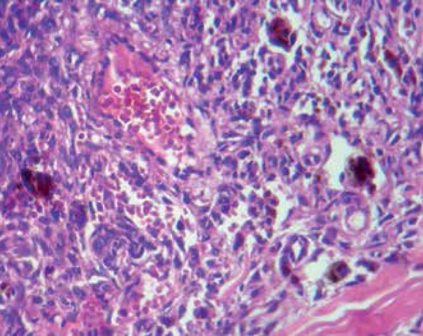
High magnification: detail of the brown pigment retention by proliferated
stellate cells. Dilated vessels filled with red blood cells in between
stellate cells (Hematoxylin & eosin x400)
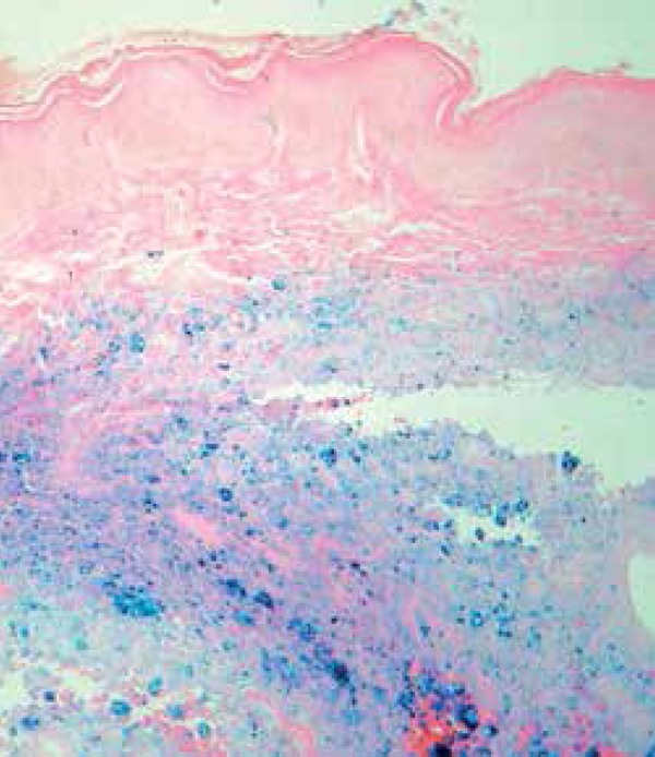
Perls’ staining revealing bluish intracellular iron storage in the form
of hemosiderin (Hematoxylin & eosin x100)
References
-
- Argenziano G, Soyer HP, De Giorgi V, Piccolo D, Carli P, Delfino M, et al. Dermoscopy: A Tutorial. Milan: EDRA Medical Publishing & New Media; 2000.
-
- Zaballos P, Llambrich A, Ara M, Olazarán Z, Malvehy J, Puig S. Dermoscopic findings of haemosiderotic and aneurysmal dermatofibroma: report of six patients. Br J Dermatol. 2006;154:244–250. - PubMed
-
- Kilinc Karaarslan I, Gencoglan G, Akalin T, Ozdemir F. Different dermoscopic faces of dermatofibromas. J Am Acad Dermatol. 2007;57:401–406. - PubMed
-
- Puig S, Romero D, Zaballos P, Malvehy J. Dermoscopy of dermatofibroma. Arch Dermatol. 2005;141:122–122. - PubMed
-
- Lourival Lopes Filho L, de Oliveira de Avelar Alchorne A. Pre-operative evaluation of the visible and palpable diameters of dermatofibroma. Int J Dermatol. 2003;42:736–737. - PubMed
Publication types
MeSH terms
LinkOut - more resources
Full Text Sources
Other Literature Sources
Medical