Review
. 2017 Apr;52(2):145-162.
doi: 10.1080/10409238.2017.1287161. Epub 2017 Feb 20.

Dynamic protein S-palmitoylation mediates parasite life cycle progression and diverse mechanisms of virulence

Affiliations
Review

Dynamic protein S-palmitoylation mediates parasite life cycle progression and diverse mechanisms of virulence

Robert W B Brown et al. Crit Rev Biochem Mol Biol. 2017 Apr.

Abstract

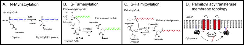
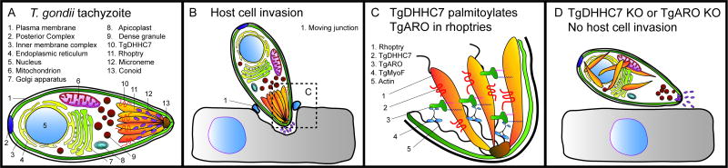
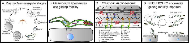
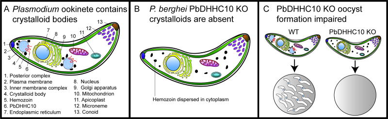
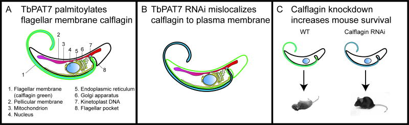
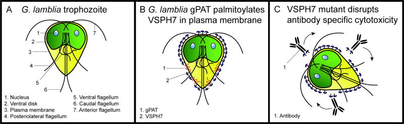
Eukaryotic parasites possess complex life cycles and utilize an assortment of molecular mechanisms to overcome physical barriers, suppress and/or bypass the host immune response, including invading host cells where they can replicate in a protected intracellular niche. Protein S-palmitoylation is a dynamic post-translational modification in which the fatty acid palmitate is covalently linked to cysteine residues on proteins by the enzyme palmitoyl acyltransferase (PAT) and can be removed by lysosomal palmitoyl-protein thioesterase (PPT) or cytosolic acyl-protein thioesterase (APT). In addition to anchoring proteins to intracellular membranes, functions of dynamic palmitoylation include - targeting proteins to specific intracellular compartments via trafficking pathways, regulating the cycling of proteins between membranes, modulating protein function and regulating protein stability. Recent studies in the eukaryotic parasites - Plasmodium falciparum, Toxoplasma gondii, Trypanosoma brucei, Cryptococcus neoformans and Giardia lamblia - have identified large families of PATs and palmitoylated proteins. Many palmitoylated proteins are important for diverse aspects of pathogenesis, including differentiation into infective life cycle stages, biogenesis and tethering of secretory organelles, assembling the machinery powering motility and targeting virulence factors to the plasma membrane. This review aims to summarize our current knowledge of palmitoylation in eukaryotic parasites, highlighting five exemplary mechanisms of parasite virulence dependent on palmitoylation.

Keywords: Giardia; Palmitoylation; Plasmodium; Toxoplasma; Trypanosoma; palmitoyl acyltransferase; pathogenesis.


Conflict of interest statement

Declaration of interest

The authors report no conflicts of interest.

Figures

Figure 1
Figure 1
Three classes of lipid modifications of proteins occur in eukaryotes: N-myristoylation, S-prenylation, and S-palmitoylation. A. N-myristoylation is catalyzed by N-myristoyltransferase (NMT) which transfers the 14-carbon saturated fatty acid myristate from myristoyl-CoA to the amino terminal glycine residue of the MGXXXS/T motif, releasing coenzyme A (CoA-SH). This reaction forms an amide bond, and is irreversible. B. S-farnesylation is catalyzed by a farnesyl protein transferase (FPTase) which transfers the 15-carbon farnesyl isoprenoid from farnesyl diphosphate to the cysteine residue of the carboxyl terminal CAAX motif (A is any aliphatic amino acid; X is one of several amino acids), releasing pyrophosphate (PPi). S-farnesylation is one of two kinds of irreversible protein S-prenylation, the other is S-geranylgeranylation which is catalyzed by geranylgeranyl protein transferase (GGTase) (not shown). C. S-palmitoylation is a reversible modification catalyzed in the forward reaction by palmitoyl acyltransferase (PAT) which transfers 16-carbon palmitate from palmitoyl-CoA to a cysteine residue in one of three types of simple motif (CXXC, XCCX, or other), releasing CoA-SH. Palmitoyl-protein thioesterase (PPT) or acyl- protein thioesterase (APT) catalyze the reverse reaction. D. Palmitoyl acyltransferase proteins typically possess 4–6 transmembrane domains (red cylinders). The canonical membrane topology is represented here with the loop containing the DHHC motif exposed in the cytoplasm, along with the amino and carboxyl termini. Two loop regions between TM1/TM2 and TM3/4 are exposed (i.e. to the outside of the cell or to the lumen of an organelle). In this panel, a myristoylated protein (left) associates with the phospholipid bilayer and is palmitoylated by a PAT, thus anchoring it firmly in the membrane (right).
Figure 2
Figure 2
The rhoptries are tethered to the actin cytoskeleton in T. gondii tachyzoites by palmitoylation of TgARO by TgDHHC7, and host cell invasion is impaired in parasites lacking either gene. A. Schematic diagram of a T. gondii tachyzoite showing its primary organelles, including three classes of secretory organelles at the apical end (micronemes, rhoptries, dense granules). TgDHHC7 with its four transmembrane domains is localized to the rhoptry membrane. B. Host cell invasion occurs when a tachyzoite attaches to the surface of the target cell, and the micronemes secrete their contents. Invasion is powered by gliding motility, and a moving junction is formed around the invading cell. The rhoptries secrete their proteins to form the parasitophorous vacuole, and the dense granules are secreted last. A tachyzoite is represented secreting its micronemes. C. The apical end of a tachyzoite contains six to fourteen rhoptries (only three are shown). TgDHHC7 is localized to the rhoptry membrane where it palmitoylates previously myristoylated TgARO, and the dual acylation firmly anchors TgARO to the rhoptry membrane. TgARO binds myosin F (TgMyoF) which is a motor protein that binds actin, thus tethering the rhoptries to the apical end of the cell. The C-terminal domain of TgARO also binds other rhoptries, clustering them together. D. Host cell invasion is impaired in TgDHHC7 or TgARO knockout cells. Palmitoylation of TgARO is necessary for its localization to the rhoptry membrane. In cells lacking either TgARO or TgDHHC7, rhoptry tethering is compromised, and the rhoptries are dispersed in the cytoplasm. Secretion of proteins from the micronemes is not affected by rhoptry dispersal in these knockouts.
Figure 3
Figure 3
Gliding motility is dependent on palmitoylation in the Plasmodium (P. berghei) insect stages. A. Life cycle of Plasmodium mosquito stages and initial invasion of host hepatocytes. The mosquito consumes a blood meal containing male and female gametocytes (not shown), which differentiate into gametes. Fertilization of the female gamete by a male gamete yields a diploid zygote, which develops into a motile ookinete that then develops into an oocyst, in which sporozoites are generated by sporogony. The sporozoites burst out of the oocyst and migrate to the salivary glands, which are injected into the human host during a blood meal. Sporozoites invade host hepatocytes to initiate infection. The liver and blood stages are not shown. B. Plasmodium zoites are the motile stages; a sporozoite is shown here adhering to the surface of a host cell and gliding. Gliding motility is powered by the glideosome (box), which is expanded in Panel C. C. The Plasmodium glideosome is shown here (top) interacting with the host cell membrane (bottom) and represents a generalized model for other apicomplexans. Microtubules stabilize the inner membrane complex (top), and contain the glideosome components GAP45 and MTIP which are palmitoylated by a PbDHHC, anchoring them to the membrane. Another major membrane protein GAP50 is not palmitoylated. GAP45 and MTIP interact with the myosin A motor protein that traverses actin filaments, generating the force which is transduced via the TRAP-like adhesin complex in the plasma membrane. The TRAP-like adhesins interact with host cell receptors in the plasma membrane during attachment. PbDHHC3 is located in the IMC. Apicomplexans possess plasma membrane DHHCs, which are probably responsible for palmitoylating these proteins. D. Gliding motility is impaired in ookinetes and sporozoites lacking PbDHHC3 or PbDHHC3/9 (double knockout). Wild type cells glide on surfaces in vitro and invade hepatocytes, whereas the gliding speed and distance is attenuated in the mutants, and they cannot invade host cells.
Figure 4
Figure 4
The crystalloid bodies form in ookinetes and are present in oocysts. A. P. berghei ookinetes are motile stages that traverse the peritrophic matrix in the mosquito midgut and epithelial cells before forming oocysts. This stage does not form a parasitophorous vacuole in the host cells so only contains micronemes, but not rhoptries or dense granules. There are 1–3 crystalloid bodies per cell, which have an unknown function, but contain proteins and lipids and possess the PAT PbDHHC10 in their membranes. The crystalloids are also surrounded by hemozoin granules. B. In the PbDHHC10 knockout parasites the crystalloids do not form, and hemozoin is dispersed in the cytoplasm. C. The wild type ookinetes normally form oocysts containing many sporozoites, but the PbDHHC10 knockout parasites form empty oocysts.
Figure 5
Figure 5
T. brucei calflagin is a flagellar membrane calcium binding protein that requires myristoylation and palmitoylation for membrane targeting and is required for virulence. A. Calflagin (green) is located in the flagellum of T. brucei parasites. The PAT that palmitoylates calflagin (TbPAT7) has been placed between Golgi and flagellar pocket; its location is currently being determined. B. Calflagin (green) is mislocalized to the pellicular (cell body) membrane upon TbPAT7 depletion by RNAi. C. Mice infected with wild type T. brucei suffer unremitting parasitemia and die within 10 days, whereas in vivo depletion of calflagin by RNAi permits the mice to survive.
Figure 6
Figure 6
G. lamblia trophozoites express variant surface proteins (VSPs) which localize to the plasma membrane, and undergo antigenic variation to mediate immune evasion. A. Schematic diagram of G. lamblia trophozoite showing its eight flagella, two nuclei, and ventral disk. B. VSPH7 is exclusively expressed on the plasma membrane surface in this trophozoite clone. VSPH7 is palmitoylated by a Giardia palmitoyl acyltransferase (gPAT) in the plasma membrane. C. A C-terminal cysteine to alanine mutant of VSPH7 did not disrupt protein localization but does lead to resistance to complement-independent antibody-mediated cytotoxicity through an unknown mechanism.

References

    1. Abrami L, Leppla SH, Van Der Goot FG. Receptor palmitoylation and ubiquitination regulate anthrax toxin endocytosis. J Cell Biol. 2006;172:309–20. - PMC - PubMed
    1. Adam RD. Biology of Giardia lamblia. Clin Microbiol Rev. 2001;14:447–75. - PMC - PubMed
    1. Aicart-Ramos C, Valero RA, Rodriguez-Crespo I. Protein palmitoylation and subcellular trafficking. Biochim Biophys Acta. 2011;1808:2981–94. - PubMed
    1. Albisetti A, Wiese S, Schneider A, Niemann M. A component of the mitochondrial outer membrane proteome of T. brucei probably contains covalent bound fatty acids. Exp Parasitol. 2015;155:49–57. - PubMed
    1. Alonso AM, Coceres VM, De Napoli MG, Nieto Guil AF, Angel SO, Corvi MM. Protein palmitoylation inhibition by 2-bromopalmitate alters gliding, host cell invasion and parasite morphology in Toxoplasma gondii. Mol Biochem Parasitol. 2012;184:39–43. - PMC - PubMed

MeSH terms

Substances

LinkOut - more resources