. 2017 Feb 20;20(2):93-99.
doi: 10.3779/j.issn.1009-3419.2017.02.10.

[Measurements of Pulmonary Artery Size for Assessment of Pulmonary Hypertension by Cardiovascular Magnetic Resonance and Clinical Application]

[Article in Chinese]
Affiliations

[Measurements of Pulmonary Artery Size for Assessment of Pulmonary Hypertension by Cardiovascular Magnetic Resonance and Clinical Application]

[Article in Chinese]
Fan Yang et al. Zhongguo Fei Ai Za Zhi. .

Abstract

Background: Pulmonary hypertension (PH) often leads to dilatation of main pulmonary artery (MPA). MPA measurements can be used to predict PH. This aim of this study is to investigate power of MPA vessel indices, which are acquired from cardiovascular magnetic resonance, to evaluate PH.

Methods: Cardiovascular-magnetic-resonance-determined parameters of MPA were acquired and calculated in 83 PH patients, whose diagnosis were confirmed with right heart catheterization and 49 healthy volunteers; these parameters included MPA diameter (DPA), ratio of DPA and ascending aorta diameter (DPA/DAo), max mean diameter (MDmax), min mean diameter (MDmin), fraction transverse diameter (fTD), fraction longitudinal diameter (fLD), and distensibility.

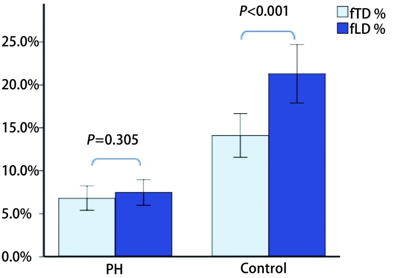
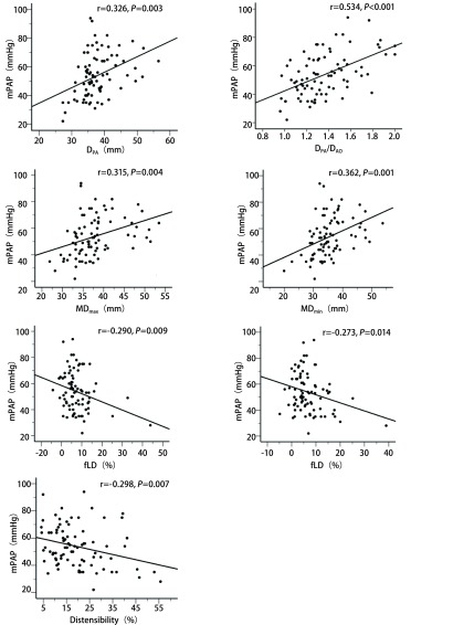
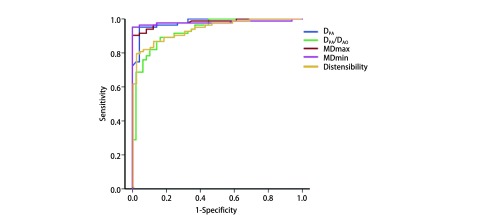
Results: Compared with control group, DPA, DPA/DAo, MDmax, and MDmin were significantly higher in patients with PH (P<0.001); fTD, fLD, and distensibility significantly decreased (P<0.001). fTD was lesser than fLD in control group (P<0.001), whereas difference was not observed in PH (P=0.305). MPA indices were significantly correlated with mean pulmonary arterial pressure (mPAP) (P<0.05), and strongest correlation was observed for DPA/DAo (r=0.534, P<0.001). By receiver operating characteristic analysis, MDmin>28.4 mm, and MDmax>32.4 mm (area under the curve, AUC=0.979, 0.981) showed best performance in predicting PH, yielding highest specificity at 100%.

Conclusions: Noninvasive cardiovascular-magnetic-resonance-derived MPA measurements provide excellent and practical reference in clinical settings for detecting PH.

背景与目的 肺高血压(pulmonary hypertension, PH)将导致主肺动脉扩张,主肺动脉(main pulmonary artery, MPA)径线测量可被临床用于评估PH。本研究旨在探讨心血管磁共振(cardiovascular magnetic resonance, CMR)测量MPA各径线参数评价PH的价值。方法 对经右心导管检查(right heart catheterization, RHC)确诊的83例PH患者及49例健康志愿者CMR图像进行分析,测量和计算主肺动脉横径(main pulmonary artery diameter, DPA)、DPA与升主动脉横径之比(the ratio of DPA and ascending aorta diameter, DPA/DAo)、最大平均直径(max mean diameter, MDmax)、最小平均直径(min mean diameter, MDmin)、横径变化分数(fraction transverse diameter, fTD)、纵径变化分数(fraction longitudinal diameter, fLD)及MPA顺应性。结果 PH组的DPA、DPA/DAo、MDmax、MDmin明显增大(P<0.001),fTD、fLD、顺应性显著减低(P<0.001)。对照组fTD小于fLD(P<0.001),但PH组两参数无明显差异(P=0.305)。MPA各径线参数均与平均肺动脉压(mean pulmonary arterial pressure, mPAP)显著相关(P<0.05),以DPA/DAo的相关性(r=0.534, P<0.001)最强。ROC曲线分析表明MDmin>28.4 mm和MDmax>32.4 mm预测PH的效能更高[曲线下面积(area under the curve, AUC=0.979, 0.981)],并且两者的特异性均达100%。结论 CMR无创性测量MPA径线评价PH具有临床实用价值。.


Figures

1
1
CMR、MPA图像采集及径线测量方法(A-D为同一PH患者)。A:CMR横轴位非门控FIESTA序列MPA横径最大的层面,分别测量DPA和DAo。B:由A图取平行于MPA走行方向定位,获得RV流出道和MPA图像。C、D:由B图于肺动脉瓣上方1.5 cm-2 cm处垂直于血流方向定位,获得MPA横截面Fast Cine PC序列单层多时相图像,于幅度图上手动勾画心动周期内各时相MPA内缘,在最大、最小截面积时相分别测量MPA横径(TD)和纵径(LD)。 The measurement methods of CMR MPA diameters (A-D from the same patient). A: DPA and DAo were measured from CMR non-gated FIESTA transversal image on the slice of the maximum MPA diameter. B: RV outflow tract with longitudinal MPA was acquired by localizing parallel to the direction of MPA in A. C, D: An image plane was prescribed perpendicular to the MPA flow direction and 1.5 cm-2 cm above the level of the pulmonary valve, and a single slice multi-phase cross-section images of MPA were obtained with Fast Cine PC sequence. Tracing the inner edge of MPA manually on each amplitude image during cardiac cycle was in order to measure the MPA TD and LD on the phases with the maximum and minimun area. CMR: cardiovascular magnetic resonance; MPA: main pulmonar y ar ter y; PH: pulmonary hypertension; FIESTA: fast imaging employing steady-state acquisition; DPA: main pulmonary artery diameter; DAo: ascending aorta diameter; RV: right ventricle; PC: phase contrast; TD: transverse diameter; LD: longitudinal diameter.
2
2
PH组与对照组间MPA径线参数比较。PH组DPA、MDmax、MDmin均显著升高。 Comparison of MPA vessel diameters between PH and control group. DPA, MDmax and MDmin were significantly increased in PH. MDmax: max mean diameter; MDmin: min mean diameter.
3
3
PH组及对照组各自fTD与fLD相比,对照组fTD小于fLD(P < 0.001),而PH组两参数间无显著性差异(P=0.305)。 Comparison between the fTD and fLD. FTD was smaller than fLD in control group, whereas there was no significant difference in PH. fTD: fraction transverse diameter; fLD: fraction longitudinal diameter.
4
4
PH组MPA各参数与mPAP间的相关性的散点图。DPA、DPA/DAo、MDmax、MDmin分别与mPAP呈显著正相关,其中DPA/DAo相关性最强;顺应性、fLD、fTD分别与mPAP呈负相关。 Scatterplots showed the relationships between MPA indices and mPAP. DPA, DPA/DAo, MDmax, and MDmin were positively correlated with mPAP respectively, and the strongest was found for DPA/DAo (r=0.534, P < 0.001). Disitensibility, fLD, and fTD had negative correlation with mPAP. MPA: main pulmonary artery; mPAP: mean pulmonary artery pressure.
5
5
ROC曲线显示MPA各参数预测PH的效能。MDmin> 28.4 mm和MDmax>32.4 mm预测PH效能较其他参数更高(AUC=0.979, 0.981)。 ROC curve showed the ability of MPA indices to predict PH. MDmin>28.4 mm and MDmax>32.4 mm performed relative better than others in predicting PH (AUC=0.979, 0.981, respectively). ROC: Receiveroperating characteristic.

Similar articles

Cited by

References

    1. Gali EN, Humbert M, Vachiery J, et al. 2015 ESC/ERS Guidelines for the diagnosis and treatment of pulmonary hypertension: the joint task force for the diagnosis and treatment of pulmonary hypertension of the european society of cardiology (ESC) and the european respiratory society (ERS): endorsed by: association for european paediatric and congenital cardiology (AEPC), international society for heart and lung transplantation (ISHLT) https://www.ncbi.nlm.nih.gov/pubmed/26320113 Eur Heart J. 2015;46(4):903–975. - PubMed
    1. Tan RT, Kuzo R, Goodman LR, et al. Utility of CT scan evaluation for predicting pulmonary hypertension in patients with parenchymal lung disease. Medical College of Wisconsin Lung Transplant Group. Chest. 1998;113(5):1250–1256. doi: 10.1378/chest.113.5.1250. - DOI - PubMed
    1. Devaraj A, Hansell DM. Computed tomography signs of pulmonary hypertension: old and new observations. Clin Radiol. 2009;64(8):751–760. doi: 10.1016/j.crad.2008.12.005. - DOI - PubMed
    1. Raymond TE, Khabbaza JE, Yadav R, et al. Significance of main pulmonary artery dilation on imaging studies. Ann Am Thorac Soc. 2014;11(10):1623–1632. doi: 10.1513/AnnalsATS.201406-253PP. - DOI - PMC - PubMed
    1. Shin S, King CS, Brown AW, et al. Pulmonary artery size as a predictor of pulmonary hypertension and outcomes in patients with chronic obstructive pulmonary disease. Respir Med. 2014;108(11):1626–1632. doi: 10.1016/j.rmed.2014.08.009. - DOI - PubMed