Clinical Trial
. 2017 Feb 21;46(2):197-204.
doi: 10.1016/j.immuni.2017.02.001.

Loss of PTEN Is Associated with Resistance to Anti-PD-1 Checkpoint Blockade Therapy in Metastatic Uterine Leiomyosarcoma

Affiliations
Clinical Trial

Loss of PTEN Is Associated with Resistance to Anti-PD-1 Checkpoint Blockade Therapy in Metastatic Uterine Leiomyosarcoma

Suzanne George et al. Immunity. .

Abstract

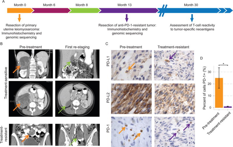
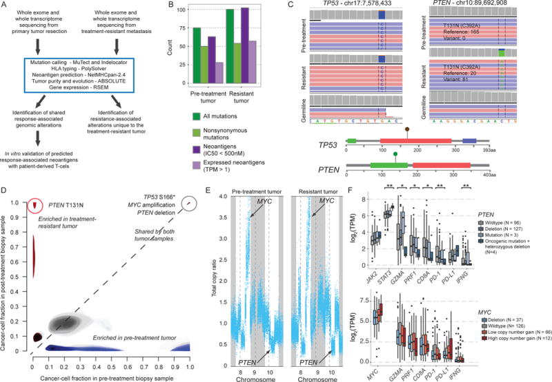
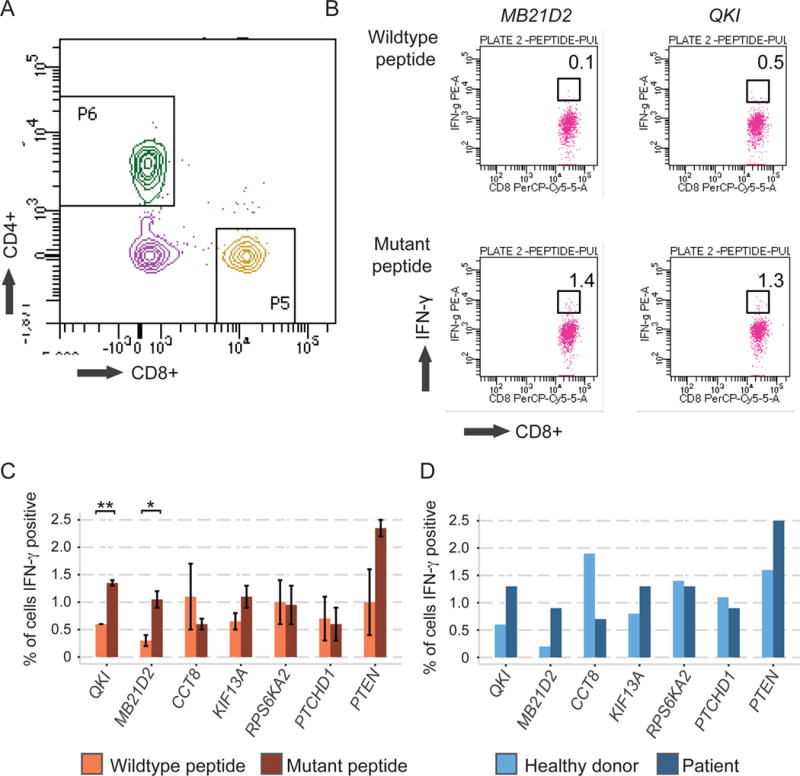
Response to immune checkpoint blockade in mesenchymal tumors is poorly characterized, but immunogenomic dissection of these cancers could inform immunotherapy mediators. We identified a treatment-naive patient who has metastatic uterine leiomyosarcoma and has experienced complete tumor remission for >2 years on anti-PD-1 (pembrolizumab) monotherapy. We analyzed the primary tumor, the sole treatment-resistant metastasis, and germline tissue to explore mechanisms of immunotherapy sensitivity and resistance. Both tumors stained diffusely for PD-L2 and showed sparse PD-L1 staining. PD-1+ cell infiltration significantly decreased in the resistant tumor (p = 0.039). Genomically, the treatment-resistant tumor uniquely harbored biallelic PTEN loss and had reduced expression of two neoantigens that demonstrated strong immunoreactivity with patient T cells in vitro, suggesting long-lasting immunological memory. In this near-complete response to PD-1 blockade in a mesenchymal tumor, we identified PTEN mutations and reduced expression of genes encoding neoantigens as potential mediators of resistance to immune checkpoint therapy.

Keywords: exceptional response; immune checkpoint; immunotherapy; neoantigen; sarcoma; whole-exome sequencing; whole-transcriptome sequencing.


Conflict of interest statement

The authors declare no conflicts of interest.

Figures

Figure 1
Figure 1. Histologic and radiographic findings in a treatment-naïve patient with metastatic uterine leiomyosarcoma receiving anti-PD-1 monotherapy
(A) Clinical course and tissue collection for immunohistochemical assessment and whole exome and whole transcriptome sequencing. (B) Computerized tomography (CT) imaging of treatment-responsive tumors and the sole treatment-resistant lesion. (C) Immunohistochemical staining of the primary and treatment-resistant tumors for PD-1, PD-L1, and PD-L2. (D) Quantification from representative tumor sections showing decrease in PD-1+ cell infiltration in the treatment-resistant lesion (p=0.039; Student’s t test). *p < 0.05.
Figure 2
Figure 2. Pre-treatment and post-treatment exomic features in uterine leiomyosarcoma in comparison to TCGA
(A) Computational workflow for whole exome and transcriptome analysis and neoantigen prediction. (B) Pre-treatment and resistant tumors had similar neoantigen and mutational loads. See also Table S1. (C) Integrated Genomics Viewer (IGV_2.3.57) (Thorvaldsdottir et al., 2013) and lollipop plots showing the TP53 S166* and PTEN T131N mutations in pre-treatment and treatment-resistant tumors. (D) Comparison of the estimated proportion of cancer cells harboring specific mutations in the pre-treatment (x-axis) versus resistant (y-axis) biopsy samples, with shared clonal mutations in the upper right (grey), mutations exclusive to the pre-treatment tumor in the lower right (blue), and mutations exclusive to the treatment-resistant tumor in the upper left (red). Lighter shading indicates mutations with cancer cell fraction distributions with high uncertainty. (E) Copy number plots show amplification of chromosome 8 and deletion of chromosome 10 affecting MYC and PTEN, respectively, in both tumors. See also Figure S1. (F) Expression of genes related to JAK/STAT and immune inhibitory signaling in untreated sarcoma tumors from the TCGA by PTEN (top) and MYC (bottom) mutational and copy number status. *p<0.05; **p<0.005
Figure 3
Figure 3. Predicted and expressed neoantigens in pre-treatment and treatment-resistant tumors
(A) Workflow of neoantigen analysis, with Venn diagram showing prioritization of putatively response-associated neoantigens. (B) Line plot showing expression of genes containing predicted neoantigens over time. Each line represents a unique neoantigen, with those synthesized for in vitro testing in green, neoantigens in MB21D2 and QKI in dark green, and all other neoantigens in gray. (C) IGV showing mRNA transcripts of mutations generating neoantigens in QKI and MB21D2 in the pre-treatment and treatment-resistant tumors. See also Figure S1, Figure S2, and Table S2.
Figure 4
Figure 4. In vitro validation of patient T cell reactivity to predicted immunogenic tumor-specific peptides
(A) Representative CD4+ versus CD8+ dot plot from viable CD3+ T cells after cell culture. (B) Representative dot plots showing proportions of interferon γ-producing CD8+ T cells following peptide incubation. (C) Bar chart showing proportions of interferon-γ-secreting CD8+ T cells following incubation with tumor-specific mutant peptides (dark orange) or corresponding wildtype control peptides (light orange). Error bars show standard error (SE) above and below the mean. (D) Comparison of reactivity of healthy donor PBMCs (light blue) to patient PBMCs (dark blue) to tumor-specific mutant peptides. See also Experimental Procedures, Supplemental Information, and Figure S4. *p<0.05; **p<0.005

References

    1. Anagnostou V, Smith KN, Forde PM, Niknafs N, Bhattacharya R, White J, Zhang T, Adleff V, Phallen J, Wali N, et al. Evolution of neoantigen landscape during immune checkpoint blockade in non-small cell lung cancer. Cancer Discovery 2017 - PMC - PubMed
    1. Barretina J, Taylor BS, Banerji S, Ramos AH, Lagos-Quintana M, Decarolis PL, Shah K, Socci ND, Weir BA, Ho A, et al. Subtype-specific genomic alterations define new targets for soft-tissue sarcoma therapy. Nat Genet. 2010;42:715–721. - PMC - PubMed
    1. Brastianos PK, Carter SL, Santagata S, Cahill DP, Taylor-Weiner A, Jones RT, Van Allen EM, Lawrence MS, Horowitz PM, Cibulskis K, et al. Genomic characterization of brain metastases reveals branched evolution and potential therapeutic targets. Cancer Discov. 2015;5:1164–1177. - PMC - PubMed
    1. Brennan CW, Verhaak RG, McKenna A, Campos B, Noushmehr H, Salama SR, Zheng S, Chakravarty D, Sanborn JZ, Berman SH, et al. The somatic genomic landscape of glioblastoma. Cell. 2013;155:462–477. - PMC - PubMed
    1. Carter SL, Cibulskis K, Helman E, McKenna A, Shen H, Zack T, Laird PW, Onofrio RC, Winckler W, Weir BA, et al. Absolute quantification of somatic DNA alterations in human cancer. Nat Biotechnol. 2012;30:413–421. - PMC - PubMed

Publication types

MeSH terms