. 2017 May 1;312(5):F897-F907.
doi: 10.1152/ajprenal.00643.2016. Epub 2017 Feb 22.

Endothelial colony-forming cells ameliorate endothelial dysfunction via secreted factors following ischemia-reperfusion injury

Affiliations

Endothelial colony-forming cells ameliorate endothelial dysfunction via secreted factors following ischemia-reperfusion injury

Jason A Collett et al. Am J Physiol Renal Physiol. .

Abstract

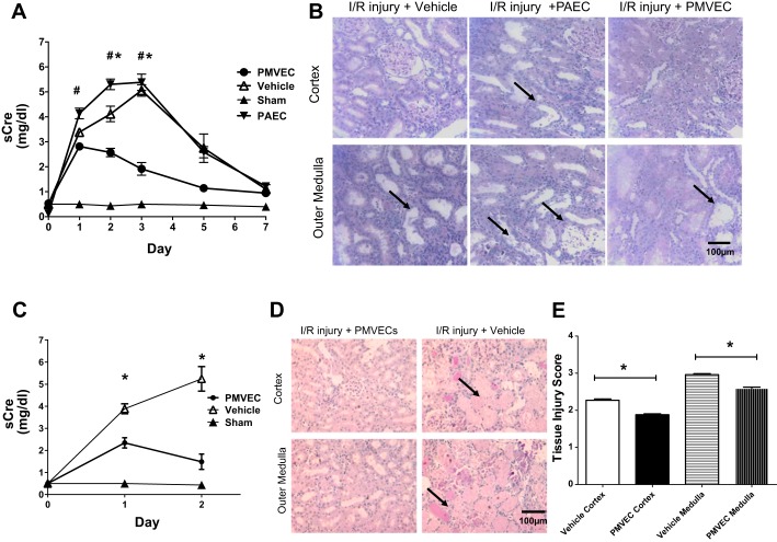
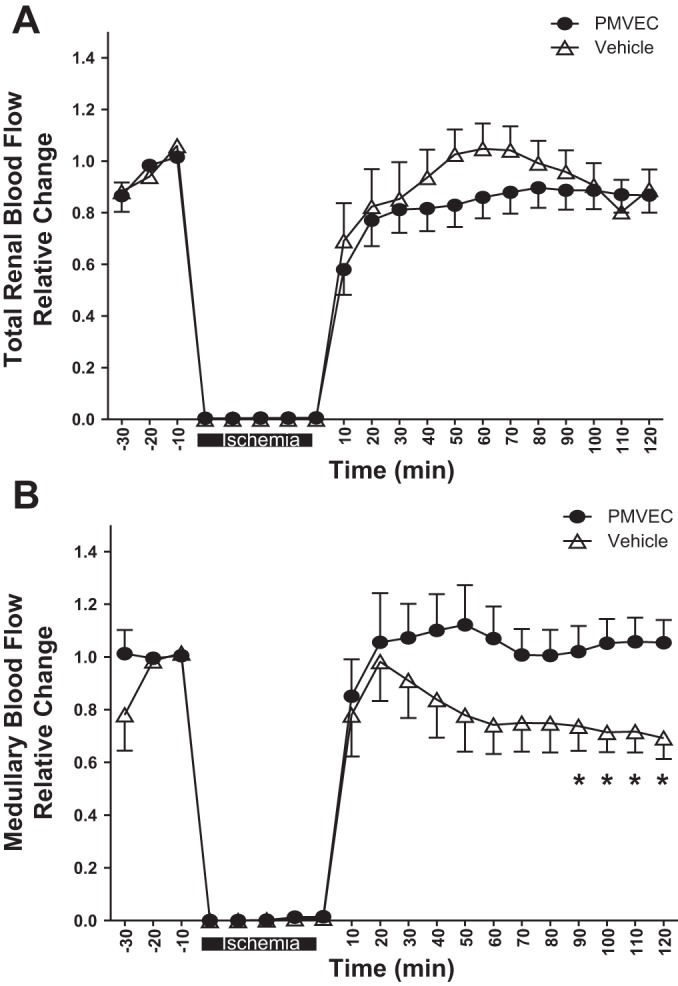
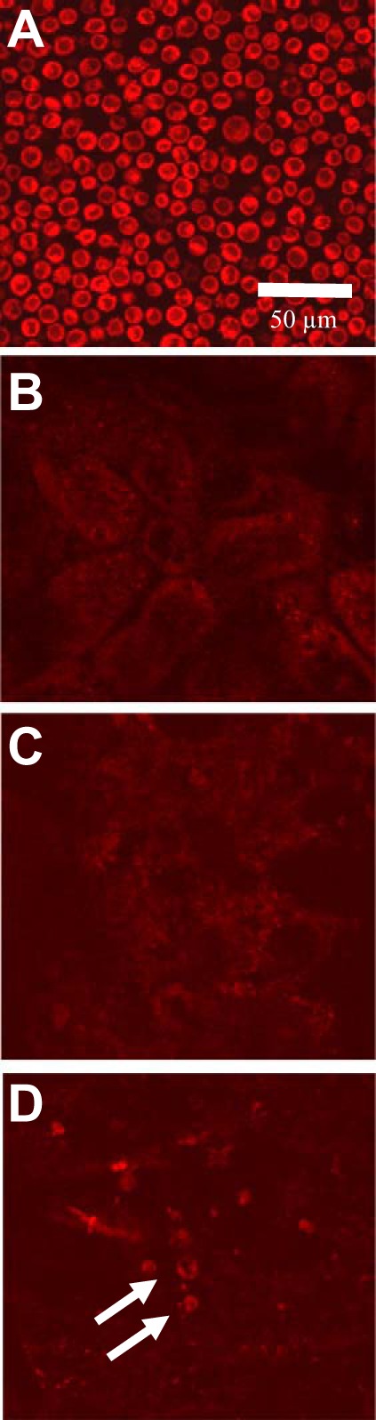
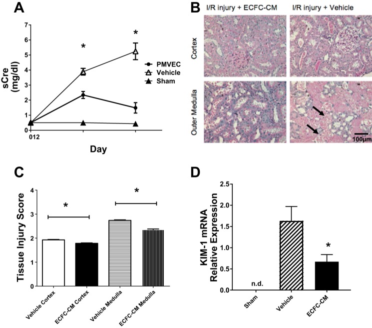
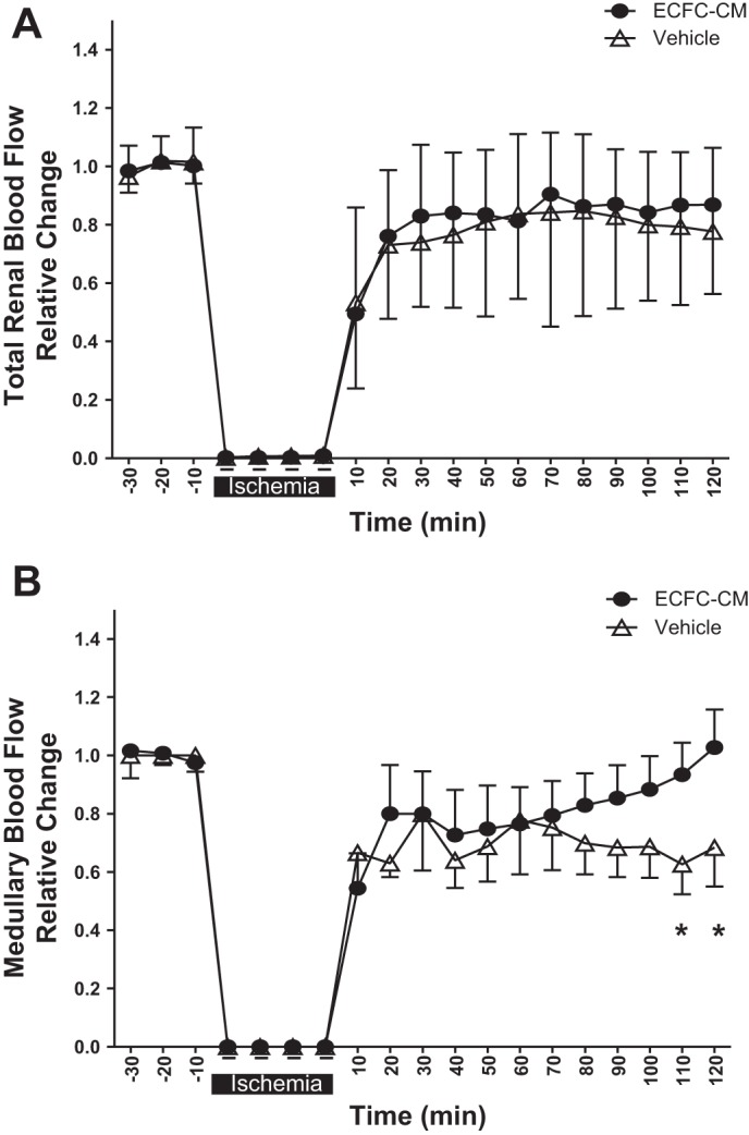
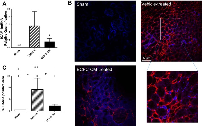
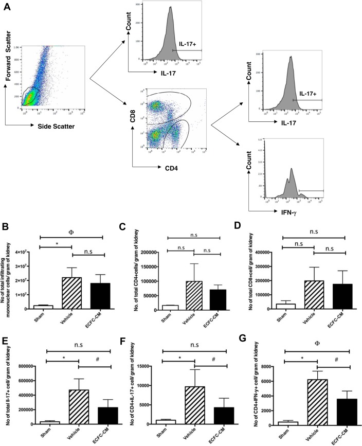
Damage to endothelial cells contributes to acute kidney injury (AKI) by leading to impaired perfusion. Endothelial colony-forming cells (ECFC) are endothelial precursor cells with high proliferative capacity, pro-angiogenic activity, and in vivo vessel forming potential. We hypothesized that ECFC may ameliorate the degree of AKI and/or promote repair of the renal vasculature following ischemia-reperfusion (I/R). Rat pulmonary microvascular endothelial cells (PMVEC) with high proliferative potential were compared with pulmonary artery endothelial cells (PAEC) with low proliferative potential in rats subjected to renal I/R. PMVEC administration reduced renal injury and hastened recovery as indicated by serum creatinine and tubular injury scores, while PAEC did not. Vehicle-treated control animals showed consistent reductions in renal medullary blood flow (MBF) within 2 h of reperfusion, while PMVEC protected against loss in MBF as measured by laser Doppler. Interestingly, PMVEC mediated protection occurred in the absence of homing to the kidney. Conditioned medium (CM) from human cultured cord blood ECFC also conveyed beneficial effects against I/R injury and loss of MBF. Moreover, ECFC-CM significantly reduced the expression of ICAM-1 and decreased the number of differentiated lymphocytes typically recruited into the kidney following renal ischemia. Taken together, these data suggest that ECFC secrete factors that preserve renal function post ischemia, in part, by preserving microvascular function.

Keywords: angiogenesis; endothelial progenitor; hemodynamics; regeneration.


Figures

Fig. 1.
Fig. 1.
Rat pulmonary microvascular endothelial cells (PMVEC) protect against renal ischemia-reperfusion (I/R) injury and accelerate functional and structural recovery. A: serum creatinine (sCre) values for 7 days following I/R or sham surgery (n = 3) in rats treated with vehicle (n = 7), pulmonary artery endothelial cells (PAEC; n = 6), or PMVEC (n = 8). B: representative photomicrographs of periodic acid-Schiff (PAS)-stained kidney sections following 7 days of recovery from renal I/R. Dilated tubules are prominent in vehicle and PAEC-treated rats (black arrows), with some evidence of cell sloughing. Magnification is shown. C: results from a 2-day time course study comparing renal function in vehicle (n = 6)-treated vs. PMVEC-treated rats (n = 6) following I/R injury. D: corresponding photomicrographs of PAS-stained kidney sections following 2 days of recovery from renal I/R, indicating a greater degree of necrotic tubules (black arrows) in vehicle-treated rats. E: injury score corresponding to 2-day postischemic rats. Data are means ± SE. * and # indicate P < 0.05 in PMVEC-treated rats compared with PAEC- and vehicle-treated rats, respectively by Student’s t-test.
Fig. 2.
Fig. 2.
Rat PMVEC preserve medullary blood flow in the early postischemic period. Total renal blood (A) and medullary blood flow (B) were monitored for 30 min before ischemia and up to 120 min postreperfusion as labeled. Data are averaged in 10-min time bins normalized to the baseline values for each rat, and data are means ± SE *P < 0.05 in vehicle- relative to PMVEC-treated rats by ANOVA with repeated measures.
Fig. 3.
Fig. 3.
Rat PMVEC do not home to the kidney following transplantation. Shown are representative confocal images of freshly suspended PMVEC labeled with cell tracker red in vitro and imaged before transplantation (A). There were no labeled cellular structures in the kidney at either 2 h (B) or 2 days (C) posttransplantation. D: fluorescently labeled cells in spleen with a similar size and fluorescent intensity of preinfused PMVEC (white arrows). Similarly, labeled structures were readily apparent in lung (not shown). Magnification is shown in A.
Fig. 4.
Fig. 4.
Human endothelial colony-forming cells-conditioned medium (ECFC-CM) protects against renal I/R injury. A: serum creatinine for 2 days following I/R or sham surgery (n = 3) in vehicle (n = 7)- and ECFC-CM-treated rats (n = 7). B: representative photomicrographs of PAS-stained kidney sections following 2 days of recovery from renal I/R. Significant necrotic debris and tubular damage are present in vehicle-treated rat kidney 2 days postsurgery (black arrows). Magnification is shown. Tissue injury score is shown in C. D: KIM-1 mRNA expression in sham, vehicle-, or ECFC-CM-treated rats. Data are means ± SE. n.d., Not detectable. *P < 0.05 in ECFC-CM-treated relative to vehicle-treated rats by Student’s t-test.
Fig. 5.
Fig. 5.
Human ECFC-CM preserves medullary blood flow in the early postischemic period. Total renal blood flow (A) and medullary blood flow (B) were monitored for 30 min before ischemia and up to 120 min postreperfusion as labeled. Data are averaged in 10-min time bins normalized to the baseline values for each rat and data are means ± SE. *P < 0.05 in vehicle- relative to ECFC-CM-treated rats by ANOVA with repeated measures.
Fig. 6.
Fig. 6.
Human ECFC-CM reduces adhesion molecular expression following recovery from I/R injury. Rats subjected to sham surgery or renal I/R and recovery for 5 h. Rats were treated with vehicle or ECFC-CM as labeled. A: mRNA expression levels for ICAM-1 derived from total RNA of whole kidney using real-time PCR. B and C: representative images of kidney ICAM-1 immunofluorescence in sham, vehicle-, and ECFC-CM-treated rats. C: quantitative analysis of % total area of ICAM-1 immunofluorescent-stained structures. Immunofluorescence data in C are % of total area compared with the mean value of sham-operated control rats. *P < 0.05 sham vs. I/R+vehicle by Student’s t-test. #P < 0.05 in I/R+vehicle- vs. I/R+ECFC-CM-treated rats.
Fig. 7.
Fig. 7.
Human ECFC-CM reduces infiltration of inflammatory cells in kidneys following I/R. Kidney resident monocytes were isolated from kidneys harvested 2 days postsurgery/treatment. A: gating strategy for FACS analysis. Lymphocytes were gated based on forward and side scatter plot. Total number of infiltrating monocytes (B), total number of CD4+ T cells (C), CD8+ T cells (D), total IL-17+ T cells (E), CD4+IL-17+ T cells (F), and IFN-γ+ cells (G) were quantified using fluorescence-activated cell sorting. Data are expressed as total number of each cell type per gram of tissue and are means ± SE. *P < 0.05 sham vs. I/R+vehicle by Student’s t-test. ϕP < 0.05 sham vs. I/R+ECFC-CM. #P < 0.05 I/R+vehicle vs. I/R+ ECFC-CM.

References

    1. Akcay A, Nguyen Q, Edelstein CL. Mediators of inflammation in acute kidney injury. Mediators Inflamm 2009: 1, 2009. doi: 10.1155/2009/137072. - DOI - PMC - PubMed
    1. Alvarado-Moreno JA, Hernandez-Lopez R, Chavez-Gonzalez A, Yoder MC, Rangel-Corona R, Isordia-Salas I, Hernandez-Juarez J, Cerbulo-Vazquez A, Gonzalez-Jimenez MA, Majluf-Cruz A. Endothelial colony-forming cells: Biological and functional abnormalities in patients with recurrent, unprovoked venous thromboembolic disease. Thromb Res 137: 157–168, 2016. doi: 10.1016/j.thromres.2015.11.005. - DOI - PubMed
    1. Alvarez DF, Huang L, King JA, ElZarrad MK, Yoder MC, Stevens T. Lung microvascular endothelium is enriched with progenitor cells that exhibit vasculogenic capacity. Am J Physiol Lung Cell Mol Physiol 294: L419–L430, 2007. doi: 10.1152/ajplung.00314.2007. - DOI - PubMed
    1. Asahara T, Masuda H, Takahashi T, Kalka C, Pastore C, Silver M, Kearne M, Magner M, Isner JM. Bone marrow origin of endothelial progenitor cells responsible for postnatal vasculogenesis in physiological and pathological neovascularization. Circ Res 85: 221–228, 1999. doi: 10.1161/01.RES.85.3.221. - DOI - PubMed
    1. Basile DP, Yoder MC. Renal endothelial dysfunction in acute kidney ischemia reperfusion injury. Cardiovasc Hematol Disord Drug Targets 14: 3–14, 2014. doi: 10.2174/1871529X1401140724093505. - DOI - PMC - PubMed

Publication types

MeSH terms

Substances