

Activation mechanism of the G protein-coupled sweet receptor heterodimer with sweeteners and allosteric agonists
- PMID: 28228527
- PMCID: PMC5347580
- DOI: 10.1073/pnas.1700001114
Activation mechanism of the G protein-coupled sweet receptor heterodimer with sweeteners and allosteric agonists
Abstract
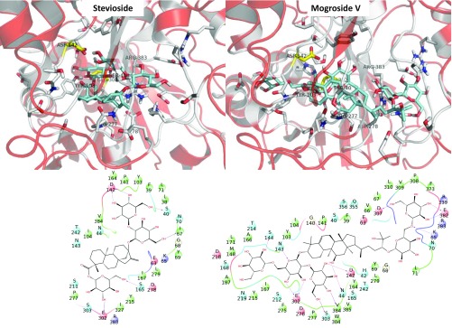
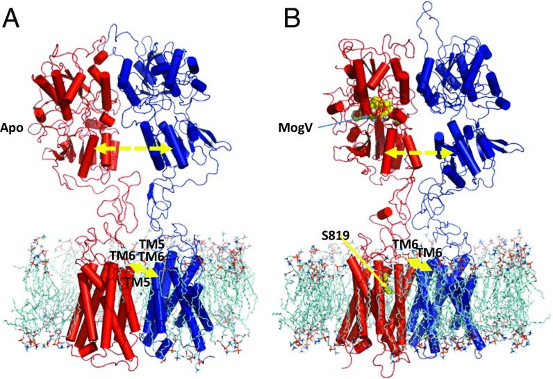
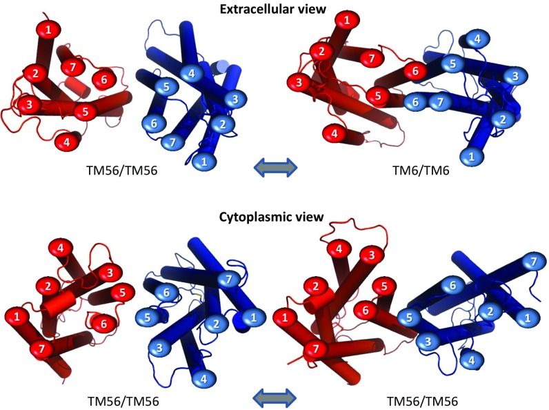
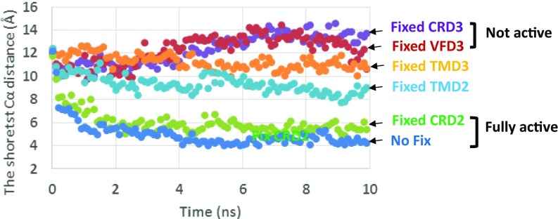
The sweet taste in humans is mediated by the TAS1R2/TAS1R3 G protein-coupled receptor (GPCR), which belongs to the class C family that also includes the metabotropic glutamate and γ-aminobutyric acid receptors. We report here the predicted 3D structure of the full-length TAS1R2/TAS1R3 heterodimer, including the Venus Flytrap Domains (VFDs) [in the closed-open (co) active conformation], the cysteine-rich domains (CRDs), and the transmembrane domains (TMDs) at the TM56/TM56 interface. We observe that binding of agonists to VFD2 of TAS1R2 leads to major conformational changes to form a TM6/TM6 interface between TMDs of TAS1R2 and TAS1R3, which is consistent with the activation process observed biophysically on the metabotropic glutamate receptor 2 homodimer. We find that the initial effect of the agonist is to pull the bottom part of VFD3/TAS1R3 toward the bottom part of VFD2/TAS1R2 by ∼6 Å and that these changes get transmitted from VFD2 of TAS1R2 (where agonists bind) through the VFD3 and the CRD3 to the TMD3 of TAS1R3 (which couples to the G protein). These structural transformations provide a detailed atomistic mechanism for the activation process in GPCR, providing insights and structural details that can now be validated through mutation experiments.
Keywords: GPCR activation; class C GPCR; molecular dynamics; noncaloric sweetener.
Conflict of interest statement
The authors declare no conflict of interest.
Figures
References
-
- Costanzi S. 2015 GPCR Structures Solved Through X-Ray Crystallography. Available at www.costanziresearch.com/p/table.html.
-
- Xue L, et al. Major ligand-induced rearrangement of the heptahelical domain interface in a GPCR dimer. Nat Chem Biol. 2015;11(2):134–140. - PubMed
-
- Maîtrepierre E, Sigoillot M, Le Pessot L, Briand L. Recombinant expression, in vitro refolding, and biophysical characterization of the N-terminal domain of T1R3 taste receptor. Protein Expr Purif. 2012;83(1):75–83. - PubMed
Publication types
MeSH terms
Substances
LinkOut - more resources
Full Text Sources
Other Literature Sources
Medical
