. 2017 Feb 13:14:14.
doi: 10.1186/s12986-017-0169-3. eCollection 2017.

Hepatic iron storage is related to body adiposity and hepatic inflammation

Affiliations

Hepatic iron storage is related to body adiposity and hepatic inflammation

Chan Yoon Park et al. Nutr Metab (Lond). .

Abstract

Background: Obesity has been reported to be associated with iron deficiency. However, few studies have investigated iron status in low adiposity. To investigate whether body adiposity was associated with altered hepatic iron status, we compared liver iron levels and markers involved in inflammation and iron absorption in obese, control, and mildly calorie restricted mice.

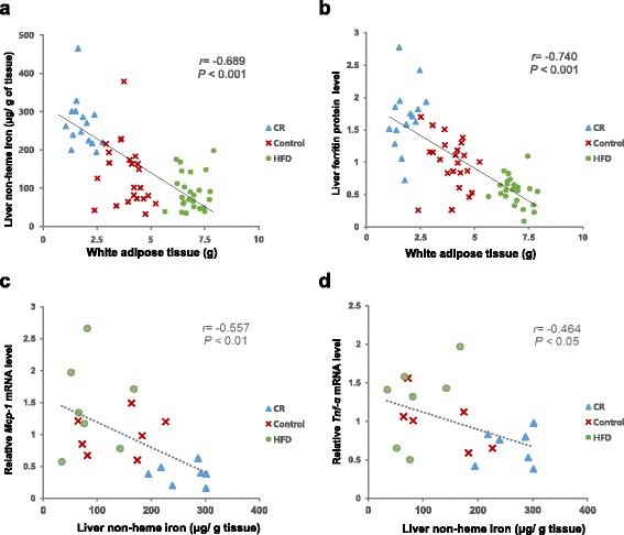
Methods: Seven week old C57BL/6 mice were fed control (10% kcal fat, Control) or high fat (60% kcal fat, HFD) diets, or reduced amount of control diet to achieve 15% calorie restriction (CR) for 16 weeks. Hepatic non-heme iron content and ferritin protein level, and hematocrit and hemoglobin levels were determined to assess iron status. Hepatic expression of Mcp-1 and Tnf-α were measured as hepatic inflammatory markers. Hepatic hepcidin (Hamp) and Bmp6, and duodenal Dmt1, Dcyt1b, hephaestin (Heph) and ferroportin mRNA levels were measured as factors involved in regulation of iron absorption.

Results: Hepatic non-heme iron and ferritin protein levels were significantly higher in the CR group compared with the Control group, and significantly lower in the HFD group. These two iron status markers showed significantly negative correlations with the amount of white adipose tissue (r = -0.689 for hepatic non-heme iron and r = -0.740 for ferritin). Hepatic Mcp-1 and Tnf-α mRNA levels were significantly lower in the CR compared with the HFD (74 and 47% lower) and showed significantly negative correlations with hepatic non-heme iron levels (Mcp-1: r = -0.557, P < 0.05; Tnf-α: r = -0.464, P < 0.05). Hepatic Hamp mRNA levels were lower in the HFD and higher in the CR groups compared with the Control group, which could be a response to maintain iron homeostasis. Duodenal Dcyt1b mRNA levels were higher in the CR group compared with the HFD group and duodenal Heph mRNA levels were higher in the CR group than the Control group.

Conclusion: We showed that body adiposity was inversely correlated with liver iron status. Low inflammation levels in hepatic milieu and enhanced expression of duodenal oxidoreductases induced by calorie restriction could have contributed to higher iron status.

Keywords: Body adiposity; Duodenal iron transporter; Hepatic non-heme iron; Hepcidin; Iron absorption; Mild calorie restriction.


Figures

Fig. 1
Fig. 1
Liver non-heme iron concentration and ferritin protein level: a Liver non-heme iron level per gram of tissue (μg/g tissue); b Densitometric analysis of liver ferritin protein expression and representative ferritin Western blots. The intensity of ferritin was densitometrically measured and normalized to the protein expression level of β-actin. abcDifferent superscript letters indicate P < 0.05. One-way ANOVA followed by Fisher’s LSD multiple comparison was used to determine significant difference. n = 16–25 per group
Fig. 2
Fig. 2
Correlations between liver iron levels and white adipose tissue or hepatic inflammatory markers: a Liver non-heme iron level (μg/g tissue) and white adipose tissue weight (g), n = 63; b Liver ferritin protein level and white adipose tissue weight (g), n = 63; c Hepatic Mcp-1 mRNA level and non-heme iron level (μg/g tissue), n = 21; d Hepatic Tnf-α mRNA level and non-heme iron level (μg/g tissue), n = 21; Pearson correlation coefficient, r, and P value are indicated for each graph
Fig. 3
Fig. 3
Relative hepatic mRNA levels of genes involved in iron homeostasis and inflammation: a Heph (n = 16–23 per group) and Bmp6 (n = 7 per group) mRNA levels; b Mcp-1 and Tnf-a. mRNA levels (n = 7 per group). Data are presented as mean ± SEM. abDifferent letters indicate P < 0.05. One-way ANOVA followed by Fisher’s LSD multiple comparison was used to determine significant difference. All values were normalized to the levels of housekeeping gene Gapdh and expressed as relative mRNA level compared to the average level of the Control group
Fig. 4
Fig. 4
The oxidation status of liver proteins: a Densitometric analysis of protein carbonyls; b Representative Western blots for protein carbonyls in the four lines of standard protein and liver protein in CR, Control, and HFD groups. The intensity of protein carbonyls was densitometrically measured and normalized by the level of standard protein. Data are presented as mean ± SEM, n = 7 for each group. One-way ANOVA followed by Fisher’s LSD multiple comparison was used to determine significant difference
Fig. 5
Fig. 5
Relative duodenal mRNA levels of ferritin and enzymes involved in iron transport: a Dmt1, Fpt(ferroportin), Heph(hephaestin), and Dcyt1b mRNA levels; b Fth1(ferritin) mRNA levels. Data are presented as mean ± SEM, n = 7–12 for each group. abDifferent letters indicate P < 0.05. One-way ANOVA followed by Fisher’s LSD multiple comparison was used to determine significant difference. All values were normalized to the levels of housekeeping gene Gapdh and expressed as relative mRNA level compared to the average level of the Control group

References

    1. Wolrd Health Organization . Micronutrient deficiencies. Iron deficiency anaemia. Wolrd Health Organization. 2015.
    1. Aeberli I, Hurrell RF, Zimmermann MB. Overweight children have higher circulating hepcidin concentrations and lower iron status but have dietary iron intakes and bioavailability comparable with normal weight children. Int J Obes. 2009;33:1111–7. doi: 10.1038/ijo.2009.146. - DOI - PubMed
    1. Chung J, Kim MS, Han SN. Diet-induced obesity leads to decreased hepatic iron storage in mice. Nutr Res. 2011;31:915–21. doi: 10.1016/j.nutres.2011.09.014. - DOI - PubMed
    1. Herter-Aeberli I, Thankachan P, Bose B, Kurpad AV. Increased risk of iron deficiency and reduced iron absorption but no difference in zinc, vitamin A or B-vitamin status in obese women in India. Eur J Nutr. 2016;55:2411–21. doi: 10.1007/s00394-015-1048-1. - DOI - PubMed
    1. Laillou A, Yakes E, Le TH, Wieringa FT, Le BM, Moench-Pfanner R, et al. Intra-individual double burden of overweight and micronutrient deficiencies among Vietnamese women. PLoS One. 2014;9:e110499. doi: 10.1371/journal.pone.0110499. - DOI - PMC - PubMed

LinkOut - more resources