Case Reports
. 2017 Jan 5;12(1):1-12.
doi: 10.1016/j.radcr.2016.12.001. eCollection 2017 Mar.

Primary neuroendocrine carcinoma of the breast: report of 2 cases and literature review

Affiliations
Case Reports

Primary neuroendocrine carcinoma of the breast: report of 2 cases and literature review

Fernando Collado-Mesa et al. Radiol Case Rep. .

Abstract

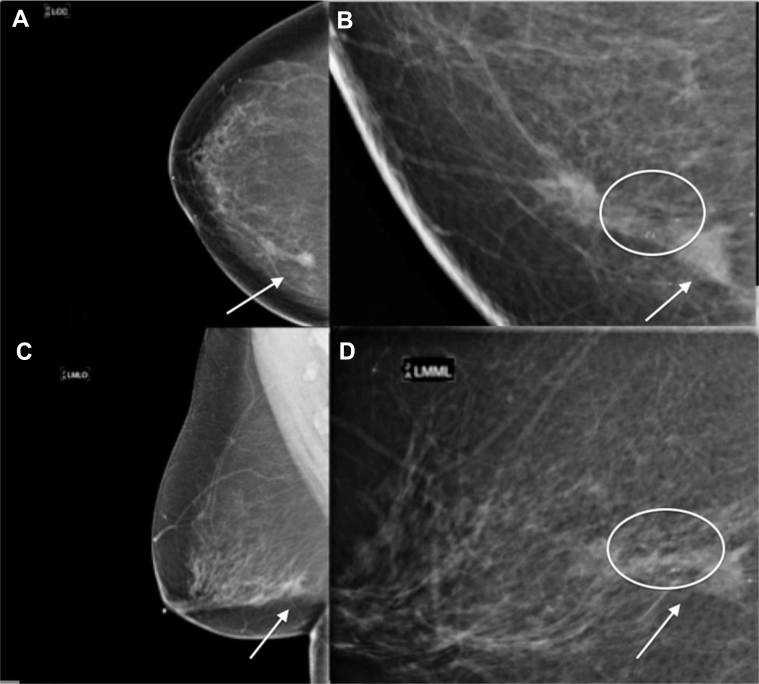
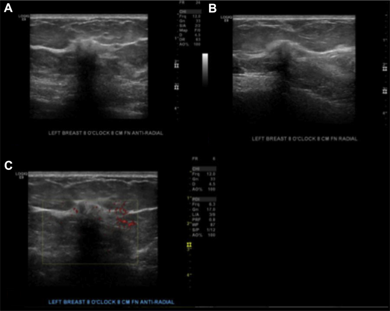
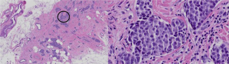
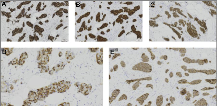
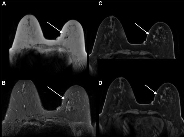
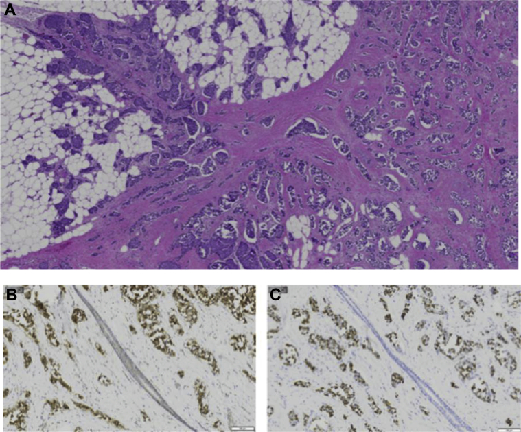
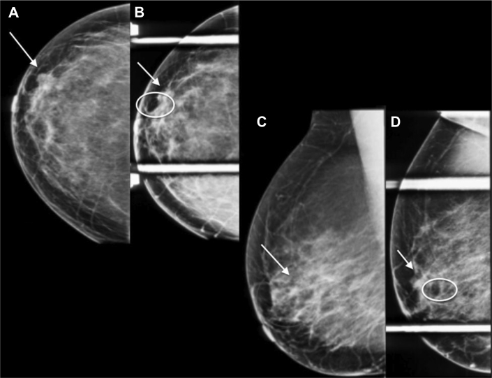
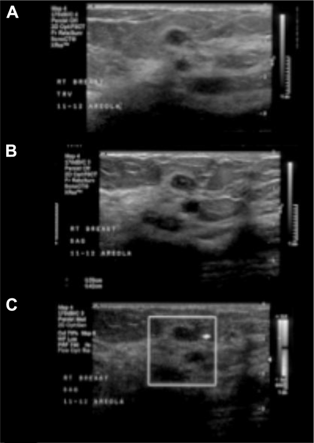
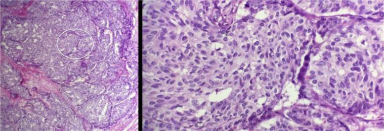
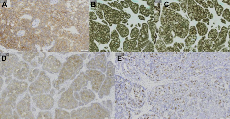
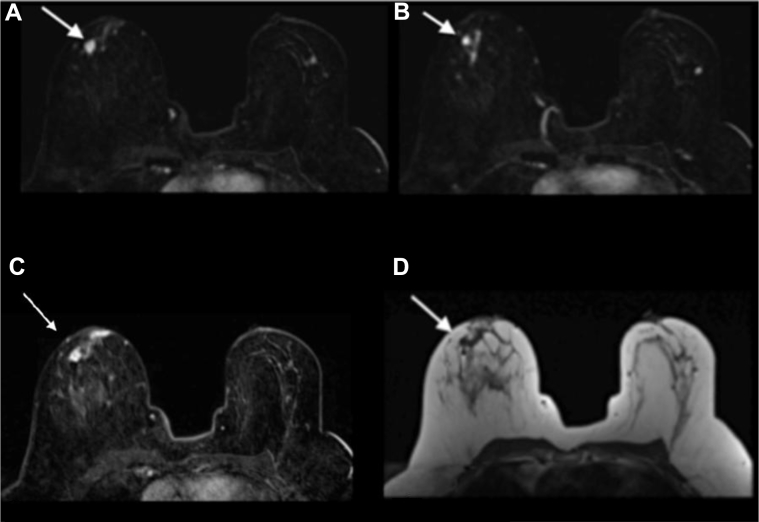
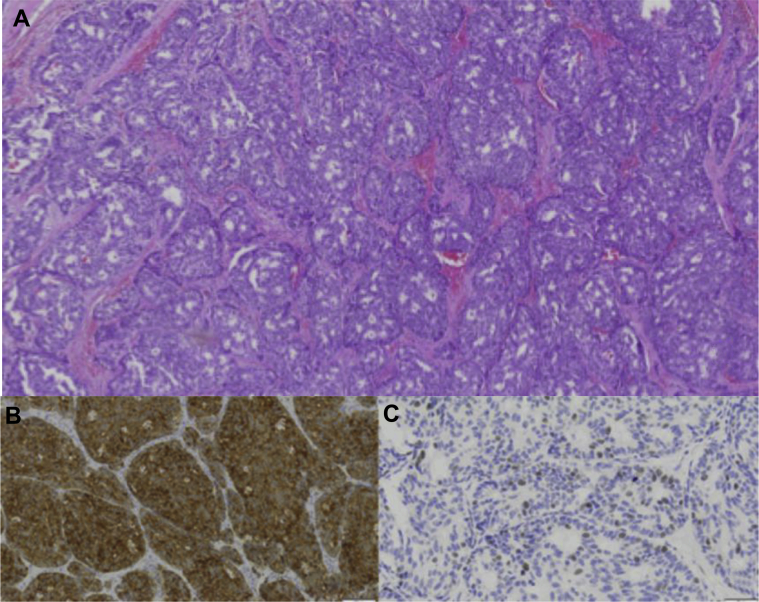
Neuroendocrine tumors of the breast are very rare accounting for less than 0.1% of all breast cancers and less than 1% of all neuroendocrine tumors. Focal neuroendocrine differentiation can be found in different histologic types of breast carcinoma including in situ and invasive ductal or invasive lobular. However, primary neuroendocrine carcinoma of the breast requires the expression of neuroendocrine markers in more than 50% of the cell population, the presence of ductal carcinoma in situ, and the absence of clinical evidence of concurrent primary neuroendocrine carcinoma of any other organ. Reports discussing the imaging characteristics of this rare carcinoma in different breast imaging modalities are scarce. We present 2 cases of primary neuroendocrine carcinoma of the breast for which mammography, ultrasound, and magnetic resonance imaging findings and pathology findings are described. A review of the medical literature on this particular topic was performed, and the results are presented.

Keywords: Breast; Carcinoma; MRI; Mammogram; Neuroendocrine; Ultrasound.


Figures

Fig. 1
Fig. 1
A 58-year-old female with left primary neuroendocrine carcinoma of the breast. Findings: left CC (A), left spot compression CC (B), left MLO (C), and left spot compression MLO (D) views demonstrate a focal asymmetry (arrow) with associated heterogeneous calcifications spanning 5 cm in length in the left breast lower inner quadrant (circle) and retroareolar heterogeneous calcifications (circle). Technique: (A) left breast full field digital mammographic craniocaudal (kVp 30; mAs 78), (B) Spot compression craniocaudal views (kVp 32; mAs 34), (C) left breast mediolateral oblique (kVp 30; mAs 80), and (D) spot compression mediolateral (kVp 32; mAs 52) projections. CC, craniocaudal; MLO, mediolateral oblique.
Fig. 2
Fig. 2
A 58-year-old female with left primary neuroendocrine carcinoma of the breast. Findings: gray scale images (A) and (B) and power Doppler images (C) show a 1.0 × 0.8 × 0.7 cm in the left breast at in the area of focal asymmetry on mammogram. Technique: (A and B) gray scale and (C) power Doppler ultrasound images of the left breast using a high-frequency linear probe.
Fig. 3
Fig. 3
A 58-year-old female with left primary neuroendocrine carcinoma of the breast. Low (left) and high (right) magnification hematoxylin and eosin stains from left breast core biopsy at 8 o’clock. Low magnification demonstrates nests and solid sheets of cells with rounded margins separated by fibrovascular stroma. High magnification demonstrates neoplastic cells which display large polygonal, granulomas, and eosinophilic cytoplasm with salt and pepper like nuclei. These pathologic findings are highly characteristic of a primary neuroendocrine carcinoma of the breast.
Fig. 4
Fig. 4
A 58-year-old female with left primary neuroendocrine carcinoma of the breast. Left breast core biopsy at 8 o’clock positive immunohistochemistry shown above include the following: (A) ER, (B) PR, (C) synaptophysin, (D) chromogranin, and (E) E-cadherin. ER, estrogen receptor; PR, progesterone receptor.
Fig. 5
Fig. 5
A 58-year-old female with left primary neuroendocrine carcinoma of the breast. Findings: (A) axial T1 precontrast image, (B) axial inversion recovery precontrast image, and (C and D) axial T1 postcontrast subtracted images of both breasts demonstrate 2 confluent spiculated masses (arrows show better) isointense on T1, heterogeneous on T2, enhance with rapid wash-in and delayed washout enhancement located in the left lower inner quadrant, in the area of suspicious mammographic and ultrasound findings. There is a biopsy clip adjacent to the most posterior mass. There are several associated adjacent small enhancing satellite lesions. The entire area of abnormality on breast MRI measures approximately 5.5 × 3.5 × 2.5 cm. Technique: breast magnetic resonance images obtained in a 3.0 Tesla magnet using a dedicated breast coil. (A) Axial MRI T1 precontrast image, TR 4.728 TE 2.292 1 mm slice thickness. (B) Axial MRI T2 stir inversion recovery precontrast image TR 5475 TE 66.816, 4 mm slice thickness. (C) Axial MRI T1 postcontrast subtracted image 1 minute, TR 6.429 TE 2.556, 2 mm slice thickness, 18-mL Prohance. (D) Axial MRI T1 postcontrast subtracted image (5 minutes), TR 6.429 TE 2.556, 2 mm slice thickness, 18 ml Prohance. MRI, magnetic resonance imaging.
Fig. 6
Fig. 6
A 58-year-old female with left primary neuroendocrine carcinoma of the breast. Left breast lumpectomy with sentinel lymph node dissection. (A) Low magnification hematoxylin and eosin stain demonstrates nests and solid sheets of cells with rounded margins separated by fibrovascular stroma. Positive immunohistochemistry shown above includes the following: (B) synaptophysin and (C) chromogranin.
Fig. 7
Fig. 7
A 62-year-old female with right primary neuroendocrine carcinoma of the breast. Findings: right CC (A), right spot compression CC (B), right MLO (C), and right spot compression MLO (D) views demonstrate a 0.9-cm oval partially obscured mass with spiculated margins (arrow) with an adjacent 0.5-cm oval circumscribed mass in the anterior right upper outer quadrant at 10 o’clock (circle). Technique: right breast digital mammogram (A) full field craniocaudal (kVp 30; mAs 72) and spot compression craniocaudal (kVp 32; mAs 38) and (B) full field mediolateral oblique (kVp 30; mAs 80) and spot compression mediolateral (kVp 32; mAs 57). CC, craniocaudal; MLO, mediolateral oblique.
Fig. 8
Fig. 8
A 62-year-old female with right primary neuroendocrine carcinoma of the breast. Findings: right breast ultrasound using a high-frequency linear probe (A and B) gray scale and (C) power Doppler images shows a 1.0 × 0.5 × 0.7 cm oval hypoechoic circumscribed parallel mass with normal sound transmission in the right breast 11–12 o'clock axes in the retroareolar region, corresponding to the largest mass on mammogram. Just deeper to this mass is a 0.6 × 0.4 × 0.8 cm oval anechoic circumscribed mass, consistent with a simple cyst, corresponding to the smallest mass on mammogram. Technique: right breast ultrasound using a high-frequency linear probe (A and B) gray scale and (C) power Doppler.
Fig. 9
Fig. 9
A 62-year-old female with right primary neuroendocrine carcinoma of the breast. Low (left) and high (right) magnification hematoxylin and eosin stains from 11 o’clock right breast core biopsy. Low magnification demonstrates nests and solid sheets of cells with rounded margins separated by fibrovascular stroma. High magnification demonstrates neoplastic cells which are uniform in shape and size with increased nuclear cytoplasmic ratio, hyperchromatic nucleus, and scant cytoplasm. These pathologic findings are highly characteristic of a primary neuroendocrine carcinoma of the breast.
Fig. 10
Fig. 10
A 62-year-old female with right primary neuroendocrine carcinoma of the breast. Right breast core biopsy at 11 o’clock positive immunohistochemistry shown above includes the following: (A) E-cadherin, (B) ER, (C) PR, (D) synaptophysin, and (E) ki67. ER, estrogen receptor; PR, progesterone receptor.
Fig. 11
Fig. 11
A 62-year-old female with right primary neuroendocrine carcinoma of the breast. Findings: (A) axial T1 dynamic 1 minute postcontrast fat-suppressed subtracted image demonstrates a 1.2 × 1.3 cm oval heterogeneously enhancing spiculated mass in the right retroareolar region (arrow), with an adjacent biopsy clip, corresponding to the largest mass on mammogram and ultrasound and to the biopsy-proven carcinoma. (B) axial T1 dynamic 1 minute postcontrast fat-suppressed subtracted image demonstrates a 3.8 cm in maximum length area of ductal clumped non–mass-like enhancement adjacent to the mass (arrow), without mammographic or sonographic correlate, corresponding to pathology-proven DCIS at lumpectomy. (C) Axial T1 dynamic 6 minute postcontrast fat-suppressed subtracted image demonstrates delayed washout of the right retroareolar 1.2 × 1.3 cm spiculated mass (arrow). (D) Axial T1 precontrast image shows the right retroareolar 1.2 × 1.3 cm spiculated mass (arrow). Technique: breast magnetic resonance images obtained in a 1.5 Tesla magnet using a dedicated breast coil: (A) axial MRI T1 postcontrast subtracted image 1 minute, TR 3.87 TE 1.05, 0.9 mm slice thickness, 18 mL Magnevist image 108/208. (B) Axial MRI T1 postcontrast subtracted image 1 minute, TR 3.87 TE 1.05, 0.9 mm slice thickness, 18 mL Magnevist image 112/208. (C) Axial MRI T1 postcontrast subtracted image 6 minutes, TR 3.87 TE 1.05, 0.9 mm slice thickness, 18 ml Magnevist. (D) Axial MRI T1 precontrast image, TR 449 TE 12 4 mm slice thickness. MRI, magnetic resonance imaging.
Fig. 12
Fig. 12
A 62-year-old female with right primary neuroendocrine carcinoma of the breast. Right breast lumpectomy with sentinel lymph node dissection. (A) Low magnification hematoxylin and eosin stain demonstrates nests and solid sheets of cells with rounded margins separated by fibrovascular stroma. Positive immunohistochemistry shown above includes the following: (B) synaptophysin and (C) ki 67.

References

    1. Ogawa H., Nishio A., Satake H., Naganawa S., Imai T., Sawaki M. Neuroendocrine tumor in the breast. Radiat Med. 2008;26(1):28–32. - PubMed
    1. Wang J., Wei B., Albarracin C., Hu J., Abraham S., Wu Y. Invasive neuroendocrine carcinoma of the breast: a population-based study from the surveillance, epidemiology and end results (SEER) database. Biomedcentral Cancer. 2014;14:147. - PMC - PubMed
    1. Murthy V., Geethamala K., Kumar B., Sudharao M. Primary neuroendocrine carcinoma of breast: a rare case report. Ann Med Health Sci Res. 2013;3(1):S35–S37. - PMC - PubMed
    1. Fernando A., Rodríguez J., Meek E., Sanchez J., Tawil M., Torregrosa L. Locally-advanced primary neuroendocrine carcinoma of the breast: case report and review of literature. World Journal of Surgical Oncology. 2013;11:128. - PMC - PubMed
    1. Ajisaka H., Maeda K., Miwa A. Breast cancer with endocrine differentiation: report of two cases showing different histologic patterns. Surg Today. 2003;22:909–912. - PubMed

Publication types

LinkOut - more resources