. 2017 Jan 30:8:2041731417691645.
doi: 10.1177/2041731417691645. eCollection 2017 Jan-Dec.

Vascularization and innervation of slits within polydimethylsiloxane sheets in the subcutaneous space of athymic nude mice

Affiliations

Vascularization and innervation of slits within polydimethylsiloxane sheets in the subcutaneous space of athymic nude mice

Rachel E Gurlin et al. J Tissue Eng. .

Abstract

Success of cell therapy in avascular sites will depend on providing sufficient blood supply to transplanted tissues. A popular strategy of providing blood supply is to embed cells within a functionalized hydrogel implanted within the host to stimulate neovascularization. However, hydrogel systems are not always amenable for removal post-transplantation; thus, it may be advantageous to implant a device that contains cells while also providing access to the circulation so retrieval is possible. Here we investigate one instance of providing access to a vessel network, a thin sheet with through-cut slits, and determine if it can be vascularized from autologous materials. We compared the effect of slit width on vascularization of a thin sheet following subcutaneous implantation into an animal model. Polydimethylsiloxane sheets with varying slit widths (approximately 150, 300, 500, or 1500 µm) were fabricated from three-dimensional printed molds. Subcutaneous implantation of sheets in immunodeficient mice revealed that smaller slit widths have evidence of angiogenesis and new tissue growth, while larger slit widths contain native mature tissue squeezing into the space. Our results show that engineered slit sheets may provide a simple approach to cell transplantation by providing a prevascularized and innervated environment.

Keywords: Subcutaneous implant; innervation; micro-stereolithographic three-dimensional printing; polydimethylsiloxane; vascularization.


Conflict of interest statement

Declaration of conflicting interests: The author(s) declared no potential conflicts of interest with respect to the research, authorship, and/or publication of this article.

Figures

Figure 1.
Figure 1.
Sheet fabrication, implantation, and explantation process. (a) A micro-stereolithographic 3D printer creates molds layer by layer. (b) Molds are filled with PDMS to create the sheet implants (c) with various slit widths. (d) Two sheets are implanted into the subcutaneous space of each athymic nude mouse. (e) At 28 days, the skin flap shows vessel infiltration around the 500 µm slit size implant, scale bar: 5 mm.
Figure 2.
Figure 2.
H&E staining reveals vascularized and innervated tissue. Samples were cut either in a vertical orientation (a) or in a horizontal orientation (b). Histological images post 28 days implantation into the subcutaneous space of athymic nude mice are shown for each slit width: (c, g, k) 150 µm, (d, h, l) 300 µm, (e, i, m) 500 µm, and (f, j, n) 1500 µm. Vertical sections at 4× magnification (c–f) are marked with a dashed rectangle, which were imaged again at 10× magnification (g–j). Arrows point to what appears to be invagination of native tissue into the slit space. Horizontal sections are shown in (k–n) at 20× magnification. Hollowed arrows point to erythrocyte-filled blood vessels. Scale bar: (c–f): 500 µm; (g–j): 200 µm; (k–n): 100 µm.
Figure 3.
Figure 3.
Immunohistochemistry detects both mature blood vessels and nerve tissue. Vertical sections of 500 µm slit samples were stained with H&E where arrows indicate (a) blood vessels or (b) nerves. The presence of (c) mature blood vessels via αSMA and (d) nerves via S100 were confirmed with immunohistochemistry of serial sections. Dashed white lines indicate the border of the material and tissue interface. Scale bar = 100 µm.
Figure 4.
Figure 4.
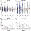
Quantification and comparison of vessel area, circularity, count, and area percentage within each slit width (150, 300, 500, 1500 µm) for both horizontal (H) and vertical (V) sections. Aggregate data of every detected vessel across all examined slit widths for (a) vessel area and (b) circularity. Each data point corresponds to a single vessel. The box plots show median (red), IQR (box), and 1.5IQR (or max and min values; whichever is shorter (whiskers)). Per slit, (c) the number of vessels per square millimeter tissue area and (d) the percentage of vessel area normalized by the total tissue area were quantified. Each data point is for a single slit. Data represent the median ± IQR. Statistical differences are indicated by ***p < 0.001, **p < 0.01, and *p < 0.05.
Figure 5.
Figure 5.
Quantification and comparison of nerve area, circularity, count, and area percentage in each slit width (150, 300, 500, 1500 µm) for both horizontal (H) and vertical (V) sections. Aggregate data of every detected nerve across all examined slit widths for (a) vessel area and (b) circularity. Each data point corresponds to a single nerve. Hollow symbols indicate that no nerves were detected in an individual slit. The box plots show the median, IQR (box), and 1.5IQR (or max and min values; whichever is shorter). Per slit, (c) the number of nerves per square millimeter tissue area and (d) the percentage of nerve area over the total tissue area were quantified. Each data point is for a single slit. Data represent the median ± IQR. Statistical differences are indicated by ***p < 0.001, **p < 0.01, and *p < 0.05.

References

    1. Langer R, Vacanti JP. Tissue engineering. Science 1993; 260(5110): 920–926. - PubMed
    1. Auger FA, Gibot L, Lacroix D. The pivotal role of vascularization in tissue engineering. Annu Rev Biomed Eng 2013; 15: 177–200. - PubMed
    1. Phelps EA, García AJ. Engineering more than a cell: vascularization strategies in tissue engineering. Curr Opin Biotechnol 2010; 21(5): 704–709. - PMC - PubMed
    1. Lovett M, Lee K, Edwards A, et al. Vascularization strategies for tissue engineering. Tissue Eng Part B Rev 2009; 15(3): 353–370. - PMC - PubMed
    1. Stosich MS, Bastian B, Marion NW, et al. Vascularized adipose tissue grafts from human mesenchymal stem cells with bioactive cues and microchannel conduits. Tissue Eng 2007; 13(12): 2881–2890. - PMC - PubMed