

The Dens: A Review of its Diverse Nomenclature and a Recommended Simplified Terminology
- PMID: 28229029
- PMCID: PMC5315575
- DOI: 10.7759/cureus.981
The Dens: A Review of its Diverse Nomenclature and a Recommended Simplified Terminology
Abstract
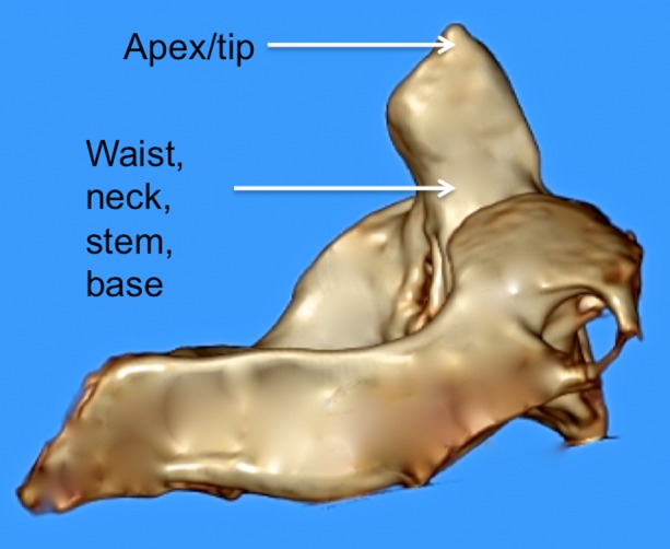
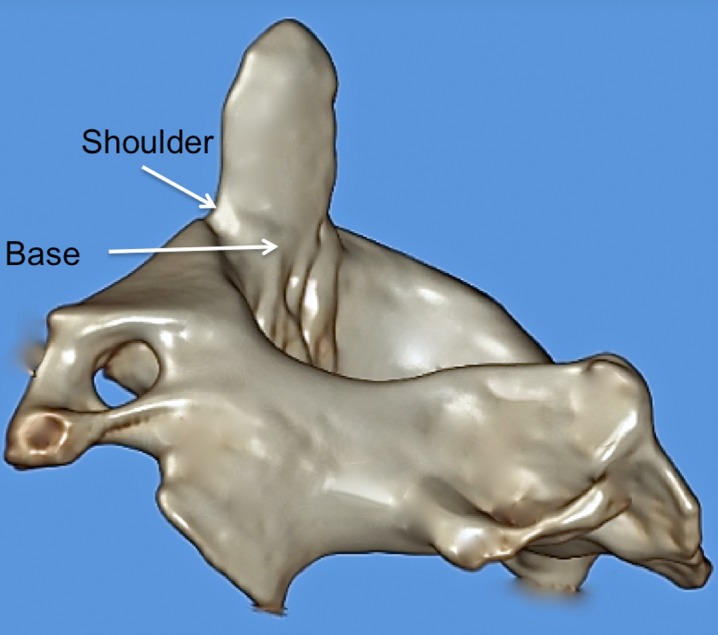
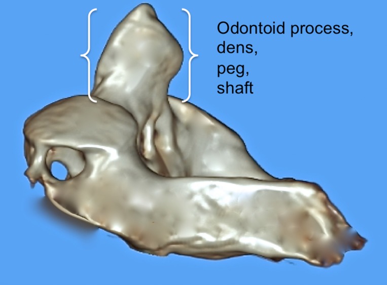
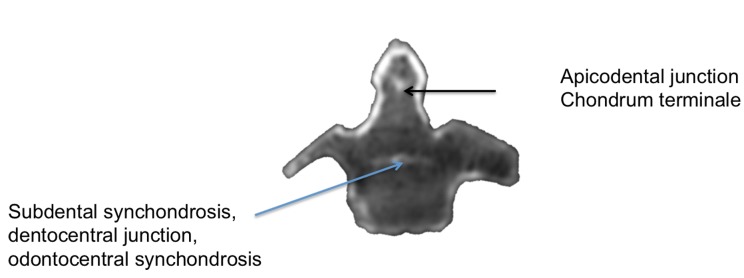
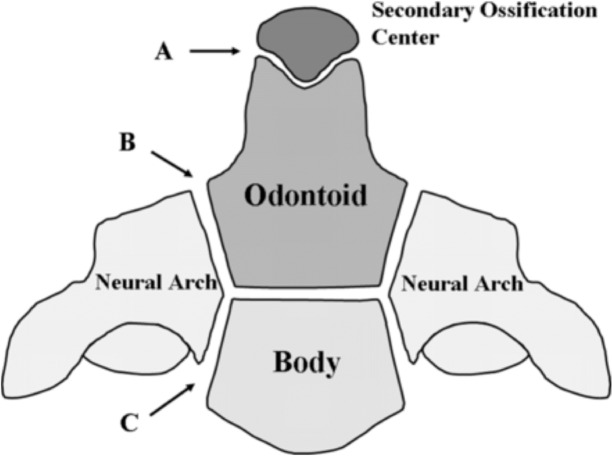
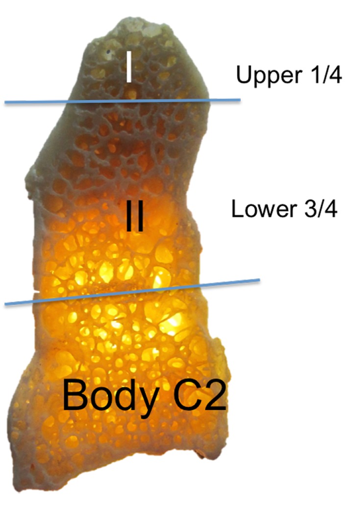
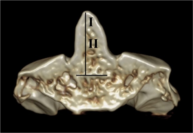
Pathology of the dens, such as fractures, demands precise terminology so that communication between physicians are succinct, diagnoses are accurate, and treatment strategies exact. This review aims to summarize the various terms used to describe the parts of the dens and recommend the ideal terminology. Using standard search engines, English language publications were searched for the many terms used to describe parts of the dens. A multitude of terms was identified with many demonstrating overlaps. Terms identified included apex, tip, apicodental, subdental, dentocentral and odontocentral junctions, peg, waist, base, neck, shaft, shoulder, and stem. Exact terminology is necessary when diagnosing or treating patients with pathology of or near the dens. The authors suggest simplified terminology for describing the parts of the dens that can be used in the future in order to be unequivocal and to avoid confusion when classifying and communicating fractures through its parts.
Keywords: anatomy; c2 vertebra; craniocervical junction; odontoid process; terminology.
Conflict of interest statement
The authors have declared that no competing interests exist.
Figures
References
-
- The odontoid process: a comprehensive review of its anatomy, embryology, and variations. Akobo S, Rizk E, Loukas M, Chapman JR, Oskouian RJ, Tubbs RS. Childs Nerv Syst. 2015;31:2025–2034. - PubMed
-
- Standring S. Edinburgh: Elsevier Churchill Livingston; 2016. Gray’s Anatomy: The Anatomical Basis of Clinical Practice. 41st Edition.
-
- Morphological and functional studies on the odontoid process of the human axis. Koebke J. Anat Embryol (Berl) 1979;155:197–208. - PubMed
-
- The three-dimensional morphometry of the odontoid peg and its impact on ventral screw osteosynthesis. Puchwein P, Jester B, Freytag B, Tanzer K, Maizen C, Gumpert R, Pichler W. Bone Joint J. 2013;95-B:536–542. - PubMed
Publication types
LinkOut - more resources
Full Text Sources
Other Literature Sources
Miscellaneous