

Bilateral Vertebral Venous Sinus Thrombosis Causing Cervical Spinal Cord Compression in a Dog
- PMID: 28229071
- PMCID: PMC5296347
- DOI: 10.3389/fvets.2017.00008
Bilateral Vertebral Venous Sinus Thrombosis Causing Cervical Spinal Cord Compression in a Dog
Abstract
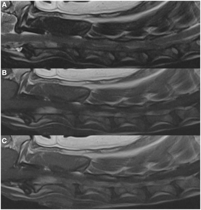
A 10-year-old male neutered mixed breed dog was evaluated for cervical hyperesthesia and tetraparesis. Magnetic resonance imaging of the brain and cervical spinal cord identified an extradural compressive lesion over the body of C2 caused by marked dilation of the vertebral venous sinuses. Following intravenous contrast administration both vertebral sinuses had heterogeneous contrast enhancement consistent with incomplete thrombi formation. An abdominal ultrasound also showed a distal aortic thrombus. A definitive cause for the thrombi formation was not identified, but the patient had several predisposing factors which may have contributed. The patient was treated with a combination of warfarin, clopidogrel, and enoxaparin as well as analgesics. Within 48 h of initiation of warfarin therapy, the tetraparesis and hyperesthesia were markedly improved. Repeat abdominal ultrasound 3 weeks after discharge showed reduction in size of aortic thrombus. Neurologic function remained normal for 6 weeks following initiation of treatment. Seventy-four days following initial diagnosis the patient rapidly declined and passed away at home. Necropsy was declined. This is the first report of vertebral venous sinus enlargement leading to spinal cord compression and tetraparesis in a dog. Additionally, warfarin in combination with clopidogrel and enoxaparin appeared to be a safe and effective treatment for the suspected thrombi reported in this case. Vertebral sinus enlargement secondary to thrombi should be considered as a differential diagnosis in patients presenting with tetraparesis and cervical hyperesthesia.
Keywords: MRI; coagulation; tetraparesis; thrombus; warfarin.
Figures


Similar articles
-
A case of suspected canine multifocal cervical venous sinus thrombosis causing cervical myelopathy.Can Vet J. 2023 Jun;64(6):534-540. Can Vet J. 2023. PMID: 37265814 Free PMC article.
-
Compressive myelopathy associated with ectasia of the vertebral and spinal arteries in a dog.Vet Pathol. 2012 Sep;49(5):779-83. doi: 10.1177/0300985811415704. Epub 2011 Aug 19. Vet Pathol. 2012. PMID: 21856870
-
[Two cases of pyogenic cervical discitis presenting tetraparesis].No Shinkei Geka. 2000 Jul;28(7):631-7. No Shinkei Geka. 2000. PMID: 10920825 Japanese.
-
Venous hypertensive myelopathy associated with cervical spondylosis.Spine J. 2016 Nov;16(11):e751-e754. doi: 10.1016/j.spinee.2016.06.003. Epub 2016 Jun 9. Spine J. 2016. PMID: 27293119 Review.
-
[Cervical myelopathy caused by bilateral vertebral artery compression].No Shinkei Geka. 1998 Jan;26(1):45-50. No Shinkei Geka. 1998. PMID: 9488991 Review. Japanese.
Cited by
-
A case of suspected canine multifocal cervical venous sinus thrombosis causing cervical myelopathy.Can Vet J. 2023 Jun;64(6):534-540. Can Vet J. 2023. PMID: 37265814 Free PMC article.
-
Morphometrical Study of the Lumbar Segment of the Internal Vertebral Venous Plexus in Dogs: A Contrast CT-Based Study.Animals (Basel). 2021 May 22;11(6):1502. doi: 10.3390/ani11061502. Animals (Basel). 2021. PMID: 34067340 Free PMC article.
References
Publication types
LinkOut - more resources
Full Text Sources
Other Literature Sources
Miscellaneous