Review
. 2016 Dec;32(6):402-410.
doi: 10.1159/000453009. Epub 2016 Dec 6.

Imaging of Cholangiocarcinoma

Affiliations
Review

Imaging of Cholangiocarcinoma

Susann-Cathrin Olthof et al. Visc Med. 2016 Dec.

Abstract

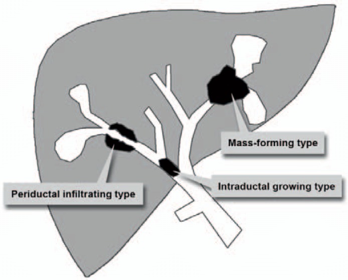
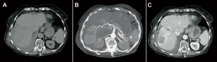
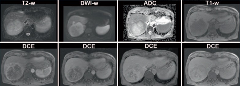
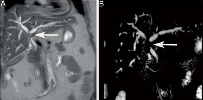
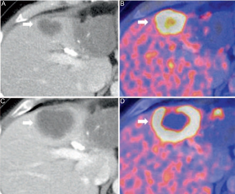
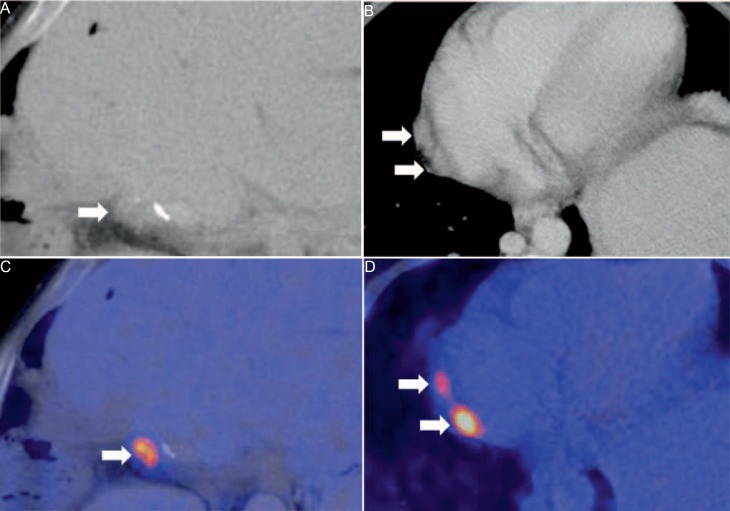
Cholangiocarcinoma (CC) is the second most common primary hepatobiliary tumour, and it is increasing in incidence. Imaging characteristics, behaviour, and therapeutic strategies in CC differ significantly, depending on the morphology and location of the tumour. In cross-sectional imaging, CCs can be classified according to the growth pattern (mass-forming, periductal infiltrating, intraductal) and the location (intrahepatic, perihilar, extrahepatic/distal). The prognosis of CC is unfavourable and surgical resection is the only curative treatment option; thus, early diagnosis (also in recurrent disease) and accurate staging including the evaluation of lymph node involvement and vascular infiltration is crucial. However, the diagnostic evaluation of CC is challenging due to the heterogeneous nature of the tumour. Diagnostic modalities used in the imaging of CC include transabdominal ultrasound, endosonography, computed tomography, magnetic resonance imaging with cholangiopancreatography, and hybrid imaging such as positron emission tomography/computed tomography. In this review, the potential of cross-sectional imaging modalities in primary staging, treatment monitoring, and detection of recurrent disease will be discussed.

Keywords: Cholangiocarcinoma; Computed tomography; Cross-sectional imaging; Magnetic resonance imaging; Positron emission tomography.


Figures

Fig. 1
Fig. 1
Schematic illustration of the three most common morphologic features of iCC according to the Liver Cancer Study Group of Japan [13].
Fig. 2
Fig. 2
CT images of an iCC. A In non-contrast enhanced images, a hypodense area is present in liver segment V-VI/VII-VIII. B Tumour vascularity can be visualized in major intensity projections of arterial-phase images. C The exact tumour size with peripheral enhancement and typical hypodense areas in the lesion centre are shown in portal-venous phase CT images.
Fig. 3
Fig. 3
MRI of a 64-year-old female patient with mass-forming CC of the right liver lobe. The CC appears hyperintense on T2-weighted images with diffusion restriction on DWI and shows a typical contrast enhancement pattern on the DCE-MRI.
Fig. 4
Fig. 4
A Coronal T2-weighted image and B MRCP of a 75-year-old female patient with periductal CC. The MRCP shows narrowing of the proximal biliary ducts (arrow) and dilation of the intrahepatic ducts.
Fig. 5
Fig. 5
18F-FDG-PET/CT for restaging of a 46-year-old male patient with iCC after chemotherapy. Hypodense iCC (A) with high tracer uptake indicating high metabolism of the lesion (B). After systemic therapy, central necrosis occurred (C) which cannot be visualized in CT alone (D).
Fig. 6
Fig. 6
45-year-old male patient. Status post hemihepatectomy for pCC 3 years ago. In CT, a small hypodense liver lesion adjacent to surgical clips is seen (A) with elevated 18F-FDG uptake (B) indicative for tumour recurrence. In addition to the local recurrence, two paracardial lymph nodes (C) show high FDG uptake in PET, which is suspicious for lymph node metastases (D).
Fig. 7
Fig. 7
49-year-old female patient after resection of pCC. In follow-up imaging, post-therapeutic changes were noticed at the resection margin (slight hyperintensity in the T2-weighted images of MRI; arrow). A Adjacent to surgical clips which are visible in CT (B). However, differentiation between local fibrotic changes and tumour recurrence was not possible based on the morphologic appearance in CT and MRI alone. In the fused 18F-FDG-PET/CT, focal 18F-FDG uptake was found in the corresponding area indicating tumour recurrence at the resection margin (C).

Similar articles

Cited by

References

    1. Patel T, Singh P. Cholangiocarcinoma: emerging approaches to a challenging cancer. Curr Opin Gastroenterol. 2007;23:317–323. - PubMed
    1. Shaib Y, El-Serag HB. The epidemiology of cholangiocarcinoma. Semin Liver Dis. 2004;24:115–125. - PubMed
    1. Khan SA, Davidson BR, Goldin R, et al. British Society of Gastroenterology Guidelines for the diagnosis and treatment of cholangiocarcinoma: consensus document. Gut. 2002;51(suppl 6):VI1–9. - PMC - PubMed
    1. Khan SA, Thomas HC, Davidson BR, Taylor-Robinson SD. Cholangiocarcinoma. Lancet. 2005;366:1303–1314. - PubMed
    1. Shaib YH, Davila JA, McGlynn K, El-Serag HB. Rising incidence of intrahepatic cholangiocarcinoma in the United States: a true increase? J Hepatol. 2004;40:472–477. - PubMed