

Characterization of the specific interaction between the DNA aptamer sgc8c and protein tyrosine kinase-7 receptors at the surface of T-cells by biosensing AFM
- PMID: 28229174
- PMCID: PMC5366180
- DOI: 10.1007/s00216-017-0238-5
Characterization of the specific interaction between the DNA aptamer sgc8c and protein tyrosine kinase-7 receptors at the surface of T-cells by biosensing AFM
Abstract
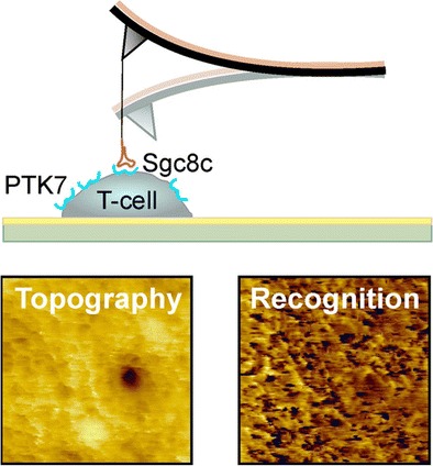
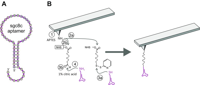
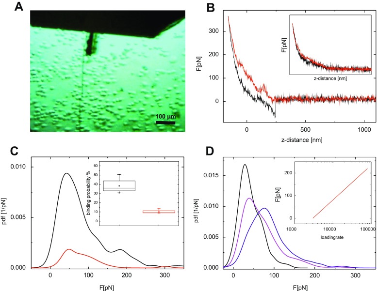
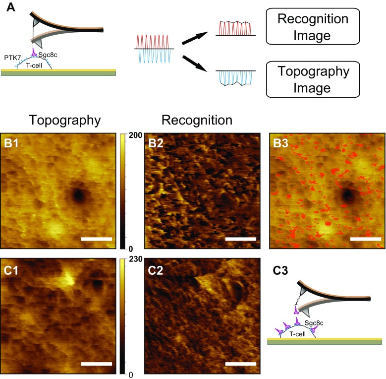
We studied the interaction of the specific DNA aptamer sgc8c immobilized at the AFM tip with its corresponding receptor, the protein tyrosine kinase-7 (PTK7) embedded in the membrane of acute lymphoblastic leukemia (ALL) cells (Jurkat T-cells). Performing single molecule force spectroscopy (SMFS) experiments, we showed that the aptamer sgc8c bound with high probability (38.3 ± 7.48%) and high specificity to PTK7, as demonstrated by receptor blocking experiments and through comparison with the binding behavior of a nonspecific aptamer. The determined kinetic off-rate (koff = 5.16 s-1) indicates low dissociation of the sgc8c-PTK7 complex. In addition to the pulling force experiments, simultaneous topography and recognition imaging (TREC) experiments using AFM tips functionalized with sgc8c aptamers were realized on the outer regions surface of surface-immobilized Jurkat cells for the first time. This allowed determination of the distribution of PTK7 without any labeling and at near physiological conditions. As a result, we could show a homogeneous distribution of PTK7 molecules on the outer regions of ALL cells with a surface density of 325 ± 12 PTK7 receptors (or small receptor clusters) per μm2. Graphical Abstract The specific interaction of the DNA aptamer sgc8c and protein tyrosine kinase-7 (PTK7) on acute lymphoblastic leukemia (ALL) cells was characterized. AFM based single molecule force spectroscopy (SMFS) yielded a kinetic off-rate of 5.16 s-1 of the complex. Simultaneous topography and recognition imaging (TREC) revealed a PTK7 density of 325 ± 12 molecules or clusters per μm2 in the cell membrane.
Keywords: DNA aptamer; Energy landscape; Molecular recognition; PTK7; Recognition imaging; Single molecule force spectroscopy; T-cell.
Conflict of interest statement
The authors declare no conflict of interest.
This article does not contain any research with human participants or animals.
Figures
Similar articles
-
Identification of the Binding Site between Aptamer sgc8c and PTK7.Anal Chem. 2024 Jul 2;96(26):10601-10611. doi: 10.1021/acs.analchem.4c01186. Epub 2024 Jun 18. Anal Chem. 2024. PMID: 38889444
-
DNA Aptamers in the Detection of Leukemia Cells by the Thickness Shear Mode Acoustics Method.Chemphyschem. 2019 Feb 18;20(4):545-554. doi: 10.1002/cphc.201801126. Epub 2019 Feb 6. Chemphyschem. 2019. PMID: 30552789
-
Aptamer-targeted DNA nanostructures with doxorubicin to treat protein tyrosine kinase 7-positive tumours.Cell Prolif. 2019 Jan;52(1):e12511. doi: 10.1111/cpr.12511. Epub 2018 Oct 12. Cell Prolif. 2019. PMID: 30311693 Free PMC article.
-
An overview of Sgc8 aptamer as a potential theranostic agent for cancer with PTK7 oncogenic target.Sci Prog. 2025 Jan-Mar;108(1):368504251325385. doi: 10.1177/00368504251325385. Sci Prog. 2025. PMID: 40033943 Free PMC article. Review.
-
Single-molecule imaging of cell surfaces using near-field nanoscopy.Acc Chem Res. 2012 Mar 20;45(3):327-36. doi: 10.1021/ar2001167. Epub 2011 Oct 12. Acc Chem Res. 2012. PMID: 21992025 Review.
Cited by
-
Aptamer-based approaches in leukemia: a paradigm shift in targeted therapy.Clin Exp Med. 2025 May 30;25(1):186. doi: 10.1007/s10238-025-01724-w. Clin Exp Med. 2025. PMID: 40445231 Free PMC article. Review.
-
Current Perspectives on Aptamers as Diagnostic Tools and Therapeutic Agents.Pharmaceutics. 2020 Jul 9;12(7):646. doi: 10.3390/pharmaceutics12070646. Pharmaceutics. 2020. PMID: 32659966 Free PMC article. Review.
-
Advances in aptamer-based nuclear imaging.Eur J Nucl Med Mol Imaging. 2022 Jul;49(8):2544-2559. doi: 10.1007/s00259-022-05782-0. Epub 2022 Apr 8. Eur J Nucl Med Mol Imaging. 2022. PMID: 35394153 Review.
-
Nanotechnology-based diagnostics and therapeutics in acute lymphoblastic leukemia: a systematic review of preclinical studies.Nanoscale Adv. 2023 Jan 11;5(3):571-595. doi: 10.1039/d2na00483f. eCollection 2023 Jan 31. Nanoscale Adv. 2023. PMID: 36756502 Free PMC article. Review.
-
Control of Ligand-Binding Specificity Using Photocleavable Linkers in AFM Force Spectroscopy.Nano Lett. 2020 May 13;20(5):4038-4042. doi: 10.1021/acs.nanolett.0c01426. Epub 2020 Apr 27. Nano Lett. 2020. PMID: 32320256 Free PMC article.
References
-
- Kawasaki ES, Clark SS, Coyne MY, Smith SD, Champlin R, Witte ON, et al. Diagnosis of chronic myeloid and acute lymphocytic leukemias by detection of leukemia-specific messenger-RNA sequences amplified in vitro. Proc Natl Acad Sci U S A. 1988;85:5698–702. doi: 10.1073/pnas.85.15.5698. - DOI - PMC - PubMed
MeSH terms
Substances
LinkOut - more resources
Full Text Sources
Other Literature Sources
Research Materials
Miscellaneous
