. 2018 Mar;55(3):1847-1860.
doi: 10.1007/s12035-017-0451-4. Epub 2017 Feb 22.

Involvement of Cellular Prion Protein in α-Synuclein Transport in Neurons

Affiliations

Involvement of Cellular Prion Protein in α-Synuclein Transport in Neurons

Laura Urrea et al. Mol Neurobiol. 2018 Mar.

Erratum in

Abstract

The cellular prion protein, encoded by the gene Prnp, has been reported to be a receptor of β-amyloid. Their interaction is mandatory for neurotoxic effects of β-amyloid oligomers. In this study, we aimed to explore whether the cellular prion protein participates in the spreading of α-synuclein. Results demonstrate that Prnp expression is not mandatory for α-synuclein spreading. However, although the pathological spreading of α-synuclein can take place in the absence of Prnp, α-synuclein expanded faster in PrPC-overexpressing mice. In addition, α-synuclein binds strongly on PrPC-expressing cells, suggesting a role in modulating the effect of α-synuclein fibrils.

Keywords: Amyloid spreading; Microfluidic devices; Prnp; Synuclein.


Conflict of interest statement

The authors declare that they have no conflict of interests.

Figures
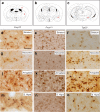
Fig. 1
Fig. 1
p-α-Synuclein pathology in the telencephalon of Prnp 0/0, Prnp +/+ and Tga20 mice injected with mouse α-synuclein fibrils in the postcommissural striatum. a-c Schemes illustrating the location of p-α-synuclein deposits (asterisks) shown in panels (do). d-o High power photomicrographs showing p-α-synuclein labelling in the striatum (df); neocortical layer V (gi); amygdala (jl) and S. nigra (mo) of Prnp 0/0 (d, g, j, m), Prnp +/+ (e, h, k, n) and Tga20 (f, i, l, o) mice. Note the relevant accumulation of p-α-synuclein labelling in intracellular deposits of retrograde-labelled projecting neurons. Scale bar: d = 100 μm pertains to eo
Fig. 2
Fig. 2
Increased α-synuclein labelling as LBL aggregates in the neocortex in Tga20 mice. ac Examples of p-α-synuclein aggregates in the motor cortex of Prnp 0/0, Prnp +/+ and Tga20 mice injected with mouse α-synuclein fibrils in the postcommissural striatum. Note the relevant accumulation of p-α-synuclein labelling in intracellular deposits of retrograde-labelled neurons in the cortical layer V of Tga20 mice. The LBL and LNL aggregates can be clearly seen over the pale background in the cortex. d Graph illustrating the quantification of the neuronal aggregates in the different genotypes. Each count represents one section. (Mice number Prnp +/+ (n = 5); Prnp 0/0 (n = 4) and Tga20 (n = 6)). In addition, the mean ± SEM is also plotted. *P < 0.05 and ***P < 0.01, ANOVA Bonferroni post hoc test. Scale bar: a = 100 μm pertains to b and c
Fig. 3
Fig. 3
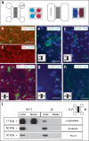
Analysis of α-synuclein transport using microfluidic devices. a 2D representation of the two PDMS devices used in the present study. b, c Double immunofluorescence photomicrographs illustrating TUJ1/GFAP (b) or TUJ1/olig2 (c) staining in primary cultured neurons in the devices. d Primary cortical cultures of the Prnp +/+ were maintained in the devices for 7 days. Then, mouse α-synuclein fibrils were added to A reservoir (asterisk), and their transport to B reservoir was analysed with immunocytochemistry (dh) and western blot (i). d Examples of double-labelled neurons (TUJ1/α-synuclein) in B reservoir (indicated with camera icon) showing α-synuclein labelling (arrows). e, f Examples of p-α-synuclein-labelled neurons and neurites (arrows) in A reservoir (indicated with camera icon). g, h Examples of p-α-synuclein-labelled neurons and axons (arrows) in B reservoir (indicated with camera icon). i Western blot showing the presence of α-synuclein (17 kDa band) in both cellular extracts (A and B) and the absence in the culture medium of B in contrast to A reservoir, avoiding passive fluid transport. Membranes were reblotted with an antibody against β-tubulin or TUJ1 for standardization and characterization. Scale bar: b, d, e, g and h = 40 μm
Fig. 4
Fig. 4
Determination of α-synuclein transport in neuronal cultures from mouse embryos carrying differing Prnp dosages. Recombinant mouse α-synuclein protofibrils were added to A reservoir (asterisk), and its binding to neurons in A reservoir as well as their transport towards B were analysed by western blot in cellular extracts. ad Examples of double-labelled Prnp 0/0 (a, b) and Prnp +/+ (c, d) neurons (TUJ1/α-synuclein) in B reservoir showing discrete cytoplasmatic α-synuclein labelling (arrows). e Examples of western blot determination of one device for each Prnp genotype for α-synuclein. Anti-β-tubulin and anti-PrPC monoclonal antibodies were used for standardization and genotype characterization. Notice that PrPC was present in WT and Tga20 cells at the time of treatment. PrPC is downregulated shortly after plating neurons, and their levels increased over time in culture (see also [68] for details). fg Densitometric analysis (see the “Material and Methods” section for details) were performed, and quantification was represented as the ratio between α-synuclein/β-tubulin detected in A reservoir after protofibril treatment (b) and ratio between α-synuclein/β-tubulin detected in B vs A reservoirs (c). Results for each device are represented by a single plot in the scatter plot. In addition, mean ± SEM is also plotted
Fig. 5
Fig. 5
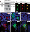
Increased binding of α-synuclein in Prnp-transfected HEK293 cells. a Western blot shows increased labelling of the 17 KDa α-synuclein band in HEK293 cells transfected with mouse Prnp-encoding plasmid in contrast to mock-transfected cells. Anti-β-tubulin monoclonal antibody was used for standardization, and anti-PrPC antibody was used to check PrPC overexpression after transfection. Notice that HEK293 cells showed a very low endogenous PrPC expression. The upper bands observed after protofibril treatment corresponded to non-monomeric forms of α-synuclein. b Examples of double-labelled GFP/α-synuclein HEK293 cells after transfection of PrPC-IRES-GFP. Note the presence of the relevant labelling in the two transfected cells in comparison to the disperse α-synuclein labelling in non-transfected cells (arrows). ce Fluorescence photomicrographs showing examples of double-labelled cells PrPC (d) overexpressing cells and α-synuclein (c). HEK293 cells were transfected with Prnp-encoding plasmid (c, d) or mock-transfected cells (e). Arrows in c and d point to double-labelled cells, and the asterisk in d labels a PrPC-positive/α-synuclein-negative HEK293 cell. Arrows in e point to α-synuclein labelling in mock-transfected cells. fh High magnification photomicrograph illustrating the distribution of α-synuclein (f) in PrPC-transfected HEK293 cells (g). Notice the relevant colocalization in several domains of the transfected cell including the plasma membrane (arrows in g and h). Scale bars: b, e = 25 μm. c, f = 25 μm belongs to d; g and h, respectively
Fig. 6
Fig. 6
a Scheme of the ΔCC, ΔHR maps and the full-length PrPC. b, c Fluorescence photomicrographs showing examples of double-labelled PrPC-synuclein cells (arrows). d, e Fluorescence photomicrographs showing examples of double-labelled cultures using the 6H4 PrPC and α-synuclein antibodies. Note the absence of double-labelled cells in these examples (arrows) and the background staining of α-synuclein labelling in non-transfected cells. hk Fluorescence photomicrographs showing examples of double-labelled ΔHR/α-synuclein cells (arrows) after treatment over the labelling of non-transfected cells. Scale bar: b, d, f, h and j = 40 μm pertains to c and e, respectively

References

    1. del Rio JA, Gavin R. Functions of the cellular prion protein, the end of Moore’s law, and Ockham’s razor theory. Prion. 2016;10(1):25–40. doi: 10.1080/19336896.2015.1126038. - DOI - PMC - PubMed
    1. Nicolas O, Gavin R, del Rio JA. New insights into cellular prion protein (PrPc) functions: the “ying and yang” of a relevant protein. Brain Res Rev. 2009;61(2):170–184. doi: 10.1016/j.brainresrev.2009.06.002. - DOI - PubMed
    1. Linden R, Martins VR, Prado MA, Cammarota M, Izquierdo I, Brentani RR. Physiology of the prion protein. Physiol Rev. 2008;88(2):673–728. doi: 10.1152/physrev.00007.2007. - DOI - PubMed
    1. Lauren J, Gimbel DA, Nygaard HB, Gilbert JW, Strittmatter SM. Cellular prion protein mediates impairment of synaptic plasticity by amyloid-beta oligomers. Nature. 2009;457(7233):1128–1132. doi: 10.1038/nature07761. - DOI - PMC - PubMed
    1. Dohler F, Sepulveda-Falla D, Krasemann S, Altmeppen H, Schluter H, Hildebrand D, Zerr I, Matschke J, Glatzel M. High molecular mass assemblies of amyloid-beta oligomers bind prion protein in patients with Alzheimer’s disease. Brain. 2014;137(Pt 3):873–886. doi: 10.1093/brain/awt375. - DOI - PubMed

Publication types