. 2017 Jun;38(6):658-668.
doi: 10.1002/humu.23201. Epub 2017 May 2.

A SAMHD1 mutation associated with Aicardi-Goutières syndrome uncouples the ability of SAMHD1 to restrict HIV-1 from its ability to downmodulate type I interferon in humans

Affiliations

A SAMHD1 mutation associated with Aicardi-Goutières syndrome uncouples the ability of SAMHD1 to restrict HIV-1 from its ability to downmodulate type I interferon in humans

Tommy E White et al. Hum Mutat. 2017 Jun.

Abstract

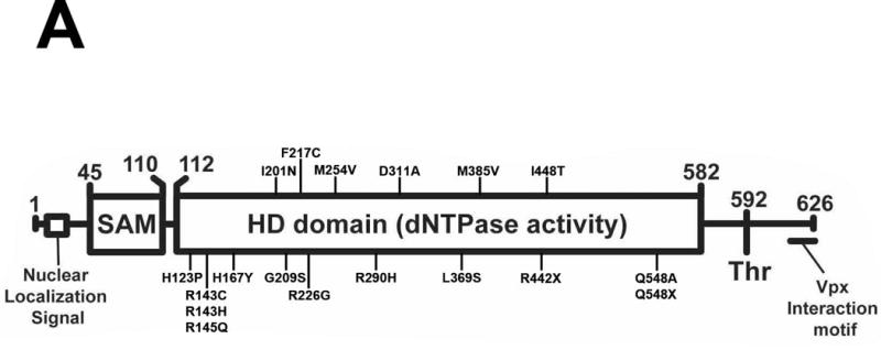
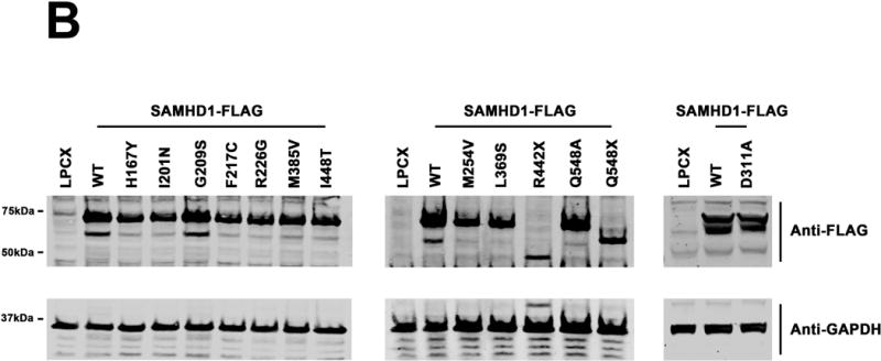
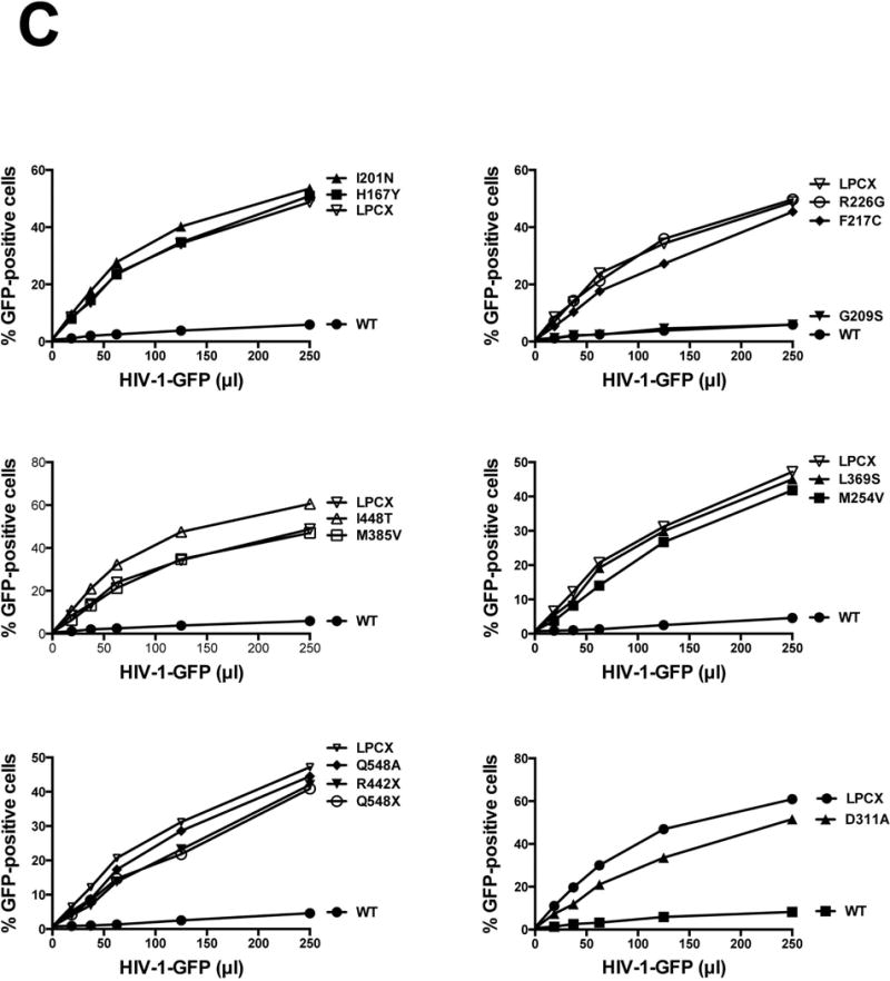
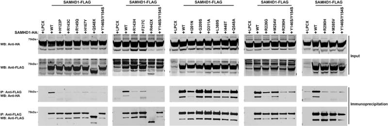
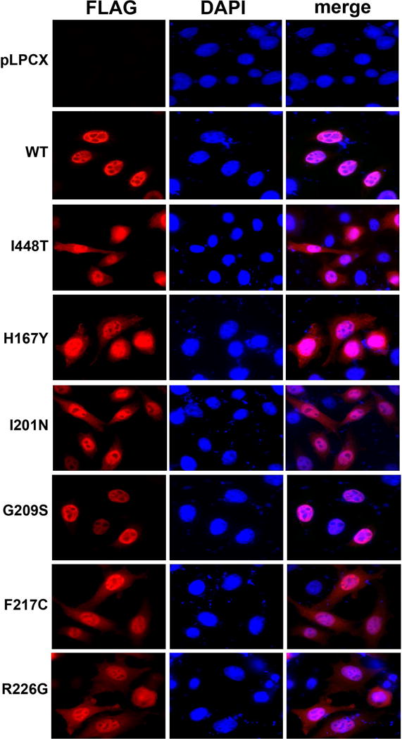
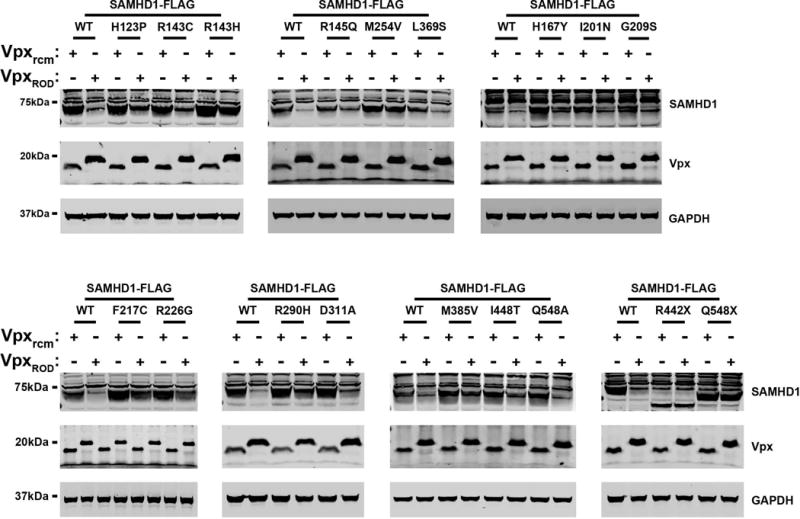
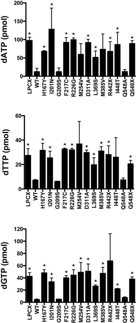
Mutations in the human SAMHD1 gene are known to correlate with the development of the Aicardi-Goutières syndrome (AGS), which is an inflammatory encephalopathy that exhibits neurological dysfunction characterized by increased production of type I interferon (IFN); this evidence has led to the concept that the SAMHD1 protein negatively regulates the type I IFN response. Additionally, the SAMHD1 protein has been shown to prevent efficient HIV-1 infection of macrophages, dendritic cells, and resting CD4+ T cells. To gain insights on the SAMHD1 molecular determinants that are responsible for the deregulated production of type I IFN, we explored the biochemical, cellular, and antiviral properties of human SAMHD1 mutants known to correlate with the development of AGS. Most of the studied SAMHD1 AGS mutants exhibit defects in the ability to oligomerize, decrease the levels of cellular deoxynucleotide triphosphates in human cells, localize exclusively to the nucleus, and restrict HIV-1 infection. At least half of the tested variants preserved the ability to be degraded by the lentiviral protein Vpx, and all of them interacted with RNA. Our investigations revealed that the SAMHD1 AGS variant p.G209S preserve all tested biochemical, cellular, and antiviral properties, suggesting that this residue is a determinant for the ability of SAMHD1 to negatively regulate the type I IFN response in human patients with AGS. Overall, our work genetically separated the ability of SAMHD1 to negatively regulate the type I IFN response from its ability to restrict HIV-1.

Keywords: Aicardi-Goutières syndrome; HIV-1; SAMHD1; interferon.


Figures

Figure 1
Figure 1. Characterization of the ability of the different SAMHD1 AGS variants to restrict HIV-1 infection. (A)
Human SAMHD1 protein is depicted showing the numbers of the amino acids residues at the boundaries of each domain along with the position and number of each AGS mutant studied here. AGS mutants studies in this work as indicates. The nuclear localization signal, the phosphorylation site on T592, and the Vpx interaction motif are depicted. Human monocytic U937 cells stably expressing the indicated mutant and wild type SAMHD1 proteins (B) were challenged with increasing amounts of HIV-1-GFP. Forthy hours post infection the percentage of GFP-positive cells was determined by flow cytometry (C). As control, U937 cells stably transduced with the empty vector LPCX were challenged with HIV-1-GFP. All SAMHD1 AGS mutant infection experiments were repeated at least 3 times and a representative figure is shown.
Figure 2
Figure 2. Oligomerization of mutant and wild type SAMHD1 proteins
Human 293T cells were cotransfected with a plasmid expressing wild type SAMHD1-HA and a plasmid either expressing wild type or mutant SAMHD1-FLAG proteins. Cells were lysed 24 hours after transfection and analyzed by Western blotting using anti-HA and anti-FLAG antibodies (Input). Subsequently, lysates were immunoprecipitated by using anti-FLAG agarose beads, as described in Materials and Methods. Anti-FLAG agarose beads were eluted using FLAG peptide, and elutions were analyzed by Western blotting using anti-HA and anti-FLAG antibodies (Immunoprecipitation). Similar results were obtained in three independent experiments and representative data is shown. WB, Western blot; IP, Immunoprecipitation; WT, wild type.
Figure 3
Figure 3. SAMHD1 AGS mutants binding to nucleic acids
Human 293T cells were transfected with plasmid expressing the indicated wild type and mutant SAMHD1 protein. Cells were lysed 24 hours after transfection (Input), were incubated with ISD-PS immobilized in StrepTactin Superflow affinity resin. Similarly, eluted proteins from the resin were visualized by Western blotting using anti-FLAG antibodies (Bound). Similar results were obtained in three independent experiments and a representative experiment is shown. ISD-PS, interferon-stimulatory DNA sequence containing a phosphorothioate backbone; WB, Western blot; WT, wild type.
Figure 4
Figure 4. Intracellular distribution of SAMHD1 AGS variant proteins in HeLa cells
HeLa cells expressing the indicated SAMHD1-FLAG variants were fixed and immunostained using antibodies against FLAG (red). Cellular nuclei were stained by using DAPI (blue). Nuclear and cytosolic distribution was quantified(Supp. Table S1).
Figure 5
Figure 5. Vpx-dependent degradation of SAMHD1 variants
HeLa cells were cotransfected with plasmids expressing the indicated SAMHD1 variants and HA-tagged Vpx from HIV-2ROD (VpxROD) or SIVRCM (Vpxrcm). Thirty-six hours post-transfection cells were harvested, and the expression levels of SAMHD1 and Vpx were analyzed by Western blotting using anti-FLAG and anti-HA antibodies. As a loading control, cell extracts were Western blotted using antibodies against GAPDH. Similar results were obtained in three independent experiments and a representative experiment is shown.
Figure 6
Figure 6. Cellular dATP, dTTP and dGTP levels in U937 cells stably expressing the different SAMHD1 variants
Quantification of dATP, dTTP and dGTP levels on PMA-treated U937 cells expressing the indicated SAMHD1 variants was performed by a primer extension assay as previously described. Similar results were obtained in three independent experiments and standard deviation is shown. “ * “ Indicates statistical significance compared to U937 cells expressing WT SAMHD1 as determined by an unpaired student t-test with P > 0.05. WT, wild type.

Similar articles

Cited by

References

    1. Ahn J, Hao C, Yan J, DeLucia M, Mehrens J, Wang C, Gronenborn AM, Skowronski J. HIV/simian immunodeficiency virus (SIV) accessory virulence factor Vpx loads the host cell restriction factor SAMHD1 onto the E3 ubiquitin ligase complex CRL4DCAF1. J Biol Chem. 2012;287(15):12550–8. - PMC - PubMed
    1. Arnold LH, Groom HC, Kunzelmann S, Schwefel D, Caswell SJ, Ordonez P, Mann MC, Rueschenbaum S, Goldstone DC, Pennell S, et al. Phospho-dependent Regulation of SAMHD1 Oligomerisation Couples Catalysis and Restriction. PLoS Pathog. 2015;11(10):e1005194. - PMC - PubMed
    1. Baldauf HM, Pan X, Erikson E, Schmidt S, Daddacha W, Burggraf M, Schenkova K, Ambiel I, Wabnitz G, Gramberg T, et al. SAMHD1 restricts HIV-1 infection in resting CD4(+) T cells. Nat Med 2012 - PMC - PubMed
    1. Behrendt R, Schumann T, Gerbaulet A, Nguyen LA, Schubert N, Alexopoulou D, Berka U, Lienenklaus S, Peschke K, Gibbert K, et al. Mouse SAMHD1 Has Antiretroviral Activity and Suppresses a Spontaneous Cell-Intrinsic Antiviral Response. Cell Rep. 2013;4(4):689–96. - PMC - PubMed
    1. Berger A, Sommer AF, Zwarg J, Hamdorf M, Welzel K, Esly N, Panitz S, Reuter A, Ramos I, Jatiani A, et al. SAMHD1-deficient CD14+ cells from individuals with Aicardi-Goutieres syndrome are highly susceptible to HIV-1 infection. PLoS Pathog. 2011;7(12):e1002425. - PMC - PubMed

Publication types

MeSH terms

Substances

Supplementary concepts