Review
. 2017 Aug:109:141-155.
doi: 10.1016/j.freeradbiomed.2017.02.010. Epub 2017 Feb 14.

Regulated methionine oxidation by monooxygenases

Affiliations
Review

Regulated methionine oxidation by monooxygenases

Bruno Manta et al. Free Radic Biol Med. 2017 Aug.

Abstract

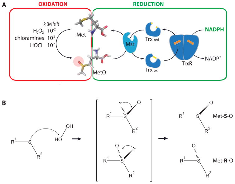
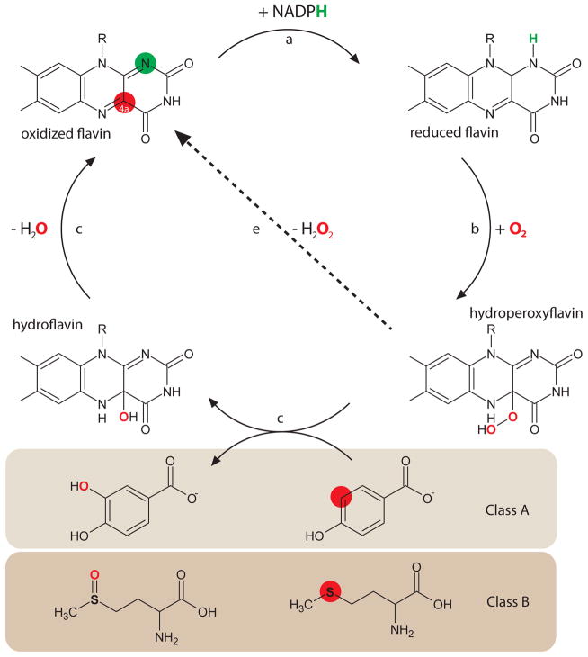
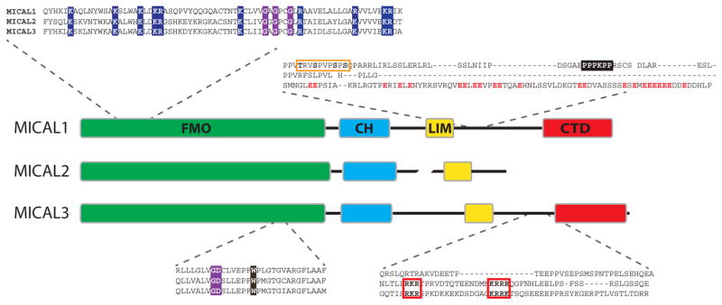
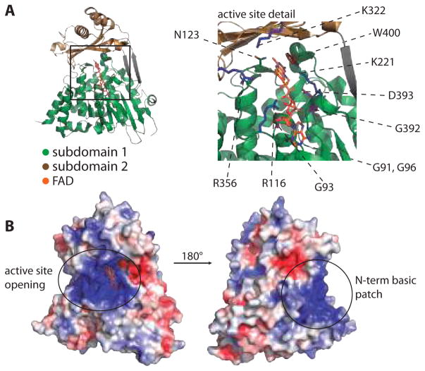
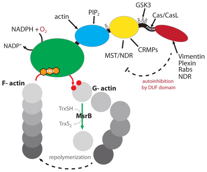
Protein function can be regulated via post-translational modifications by numerous enzymatic and non-enzymatic mechanisms, including oxidation of cysteine and methionine residues. Redox-dependent regulatory mechanisms have been identified for nearly every cellular process, but the major paradigm has been that cellular components are oxidized (damaged) by reactive oxygen species (ROS) in a relatively unspecific way, and then reduced (repaired) by designated reductases. While this scheme may work with cysteine, it cannot be ascribed to other residues, such as methionine, whose reaction with ROS is too slow to be biologically relevant. However, methionine is clearly oxidized in vivo and enzymes for its stereoselective reduction are present in all three domains of life. Here, we revisit the chemistry and biology of methionine oxidation, with emphasis on its generation by enzymes from the monooxygenase family. Particular attention is placed on MICALs, a recently discovered family of proteins that harbor an unusual flavin-monooxygenase domain with an NADPH-dependent methionine sulfoxidase activity. Based on structural and kinetic information we provide a rational framework to explain MICAL mechanism, inhibition, and regulation. Methionine residues that are targeted by MICALs are reduced back by methionine sulfoxide reductases, suggesting that reversible methionine oxidation may be a general mechanism analogous to the regulation by phosphorylation by kinases/phosphatases. The identification of new enzymes that catalyze the oxidation of methionine will open a new area of research at the forefront of redox signaling.

Keywords: Actin; DUF3585; MICAL; Methionine; Methionine oxidation; Methionine sulfoxide; Methionine sulfoxide reductase; Monooxygenase; Multidomain proteins; Redox signaling; Scaffolding proteins.


Figures

Figure 1
Figure 1. Methionine oxidation
A. Schematic representation of the chemical reaction that leads to methionine oxidation and the NAPDH-dependent reduction system based on Msr, thioredoxin (Trx) and thioredoxin reductase (TR). B. Reaction mechanism of a thioether with hydrogen peroxide. The tetrahedral intermediate is chiral, leading to chiral products, represented here as R and S enantiomers of methionine. The figure based on information reported in ,,,,,,,,,.
Figure 2
Figure 2. Reaction mechanism of flavin-dependent monooxygenases
Oxidized FAD (represented here by its isoalloxaxine ring) reacts with NAPDH forming the neutral hydroquinone (a). Molecular oxygen reacts at position C4a (red spot) forming the hydroperoxyflavin (b), that can oxidize/hydroxilate substrates (c and d) or decompose releasing H2O2 (e, “uncoupled” reaction). Class A FMOs are considered hydroxylases, as depicted here in the reaction of hydroxylation of para-hydroxybenzoate. Class B FMOs may oxidize thioether, as shown in the lower part with methionine as substrate. Adapted from,.
Figure 3
Figure 3. Domains in MICAL proteins
The domain organization of mammalian MICAL1–3 is schematically represented. Each domain is shown in a different color, conserved along the text: green for FMO, light blue for CH, yellow for LIM and red for CTD. The length of each domain is proportional to the number of amino acids in it, so do linkers, unless braked. Inserts show sequence details of mouse MICAL1–3 (NP_612188.1, NP_001180234.1 and NP_001257404.1, respectively). The conserved basic residues in the N-terminal region of the FMO domain are depicted with blue background and the GxGxxG and GD motifs with purple background. The conserved W is shown over dark brown background. Part of the connecting sequence between LIM and CTD domains is highlighted in the upper part, indicating a tandem of phosphorylation sites on MICAL1 (orange box) and the PPPKPP motif (black background), and the enrichment of acidic residues on MICAL3 (red letters). In the lower panel, the bipartite nuclear localization signal present in MICAL2 and 3 is shown with red boxes and bold letters.
Figure 4
Figure 4. Structure and conserved residues of the FMO domain
A. FMO domain of mouse MICAL1 is represented as green and brown ribbons and the FAD molecule as orange sticks, emphasizing the position of the FAD and active site at the interface between subdomains 1 and 2. A zoomed detail of the conserved residues around FAD is shown on the right side (see text for details). B. Electrostatic surface of MICAL1 FMO. The left image is shown in the same orientation as in A, while the right image is turned 180°. Positive areas are shown in blue, negative areas in red, and neutral areas in gray. Figures were done with PyMOL using PDB 2BRY and based on,,.
Figure 5
Figure 5. Structures of CH and LIM domains
A. Solution structure of human MICAL1 CH domain (PDB 2DK9) is shown as ribbons, highlighting the actin-binding region (cyan) and PIP2-binding region (pink) with sticks. The sequences of the N-terminal region of CH domains from mouse MICAL1–3 (see Figure 3) are detailed, mapping the conserved regions shown in the structure and the hydrophobic signature residues (bold). B. Solution structure of human MICAL1 LIM domain (PDB 2CO8) is shown as yellow ribbons, with cysteines and histidine involved in zinc atoms ligation (cyan spheres) represented as sticks. Figures is prepared with PyMOL, based on,.
Figure 6
Figure 6. Conservation and structure of DUF3585 domains
A. Alignment of the DUF3585 domains of mouse MICAL1 (NP-612188.1), MICAL3 (NP001257404.1), MICAL-L1 (NP803412.1), MICAL-L2 (NP777275.2), EHBP1 (NP001239444.1), EHBPL1 (NP001108069.1), EBITEIN1 (NP081863.2) and MINP (NP653101.1). Conserved residues are highlighted with red (acidic), blue (basic) or grey (hydrophobic) backgrounds. Full conservation is indicated with plain colors, while translucent colors indicated physicochemical conservation. B. The ribbon structure of human MICAL1 DUF3585 domain (PBD 5LPN) is shown on the left side and its electrostatic surface on the right. Conserved residues (numbered accordingly to mouse MICAL1) are highlighted as stick, also indicated on part A (upper line). The conserved acidic patch is encircled. Figure was prepared with PyMOL.
Figure 7
Figure 7. Proposed mechanism of MICAL
Detailed view of the FAD cofactor in oxidized (left) and reduced (right) mouse MICAL1. Colors are as in Figure 4. The conserved N123 is shown in sticks, with lateral chain atoms colored by elements (red for oxygen, blue for nitrogen). The location of C4a position is marked with a red spot. Lower panel shows the surface around FAD in the oxidized (left) and reduced (right) MICAL1, in the same orientation as in Figure 4. A 180° turn is applied to observe both sides of the protein surface. Reduction (a) produces a major conformational change that opens a channel to the C4a position. Reaction with oxygen (b) leads to the hydroperoxyflavin. Actin binds to reduced MICAL (c), and the oxidation proceeds by two alternative pathways. The position of the D-loop (detailed) can favor/induce the release of H2O2 (d1), leading to methionine oxidation by the nucleophilic attack of the sulfur on the peroxo bond (d2). Alternatively, methionine thioether can attack directly on the hydroperoxyflavin (e1), releasing hydroflavin and oxidized methionine (e2). Figure was prepared with PyMOL using PDBs 2BRY for oxidized and 2C4C for reduced mouse MICAL1 FMO domain.
Figure 8
Figure 8. F-actin structure and the role of the D-loop
Three protomers of F-actin are shown in ribbons with grey surface. Details of the D-loop connecting protomers 1 and 2 are shown in the lower panel, with methionines in stick (colored by elements, red for oxygen, yellow for sulfur). M46 is buried, while M49 is exposed to protein surface. Conservation of the D-loop methionines among 485 actin sequences from eukaryotes is shown on the right as a logo. Figure was prepared with PyMOL based on PDB 3J8A
Figure 9
Figure 9. MICALs as redox hubs
Several signals converge on MICAL architecture as shown. Integration on these signals on this multidomain protein will determine, ultimately, its activation. Once activated, MICAL proteins target actin cytoskeleton.

References

    1. Bretón-Romero R, Lamas S. Hydrogen peroxide signaling in vascular endothelial cells. Redox Biology. 2014;2:529–534. - PMC - PubMed
    1. Flohé L. Methods in Enzymology. 2010;473:1–39. - PubMed
    1. Rhee SG, et al. Intracellular messenger function of hydrogen peroxide and its regulation by peroxiredoxins. Current Opinion in Cell Biology. 2005;17:183–189. - PubMed
    1. Bak DW, et al. Cysteine-mediated redox signalling in the mitochondria. Mol Bio Syst. 2015;11:678–697. - PubMed
    1. Leichert LI, Dick TP. Incidence and physiological relevance of protein thiol switches. Biological Chemistry. 2015;396:389–399. - PubMed

MeSH terms

LinkOut - more resources