. 2017 Feb 23:7:42984.
doi: 10.1038/srep42984.

α-synuclein transfer through tunneling nanotubes occurs in SH-SY5Y cells and primary brain pericytes from Parkinson's disease patients

Affiliations

α-synuclein transfer through tunneling nanotubes occurs in SH-SY5Y cells and primary brain pericytes from Parkinson's disease patients

Birger Victor Dieriks et al. Sci Rep. .

Abstract

Parkinson's disease (PD) is characterized by the presence of inclusions known as Lewy bodies, which mainly consist of α-synuclein (α-syn) aggregates. There is growing evidence that α-syn self-propagates in non-neuronal cells, thereby contributing to the progression and spread of PD pathology in the brain. Tunneling nanotubes (TNTs) are long, thin, F-actin-based membranous channels that connect cells and have been proposed to act as conduits for α-syn transfer between cells. SH-SY5Y cells and primary human brain pericytes, derived from postmortem PD brains, frequently form TNTs that allow α-syn transfer and long-distance electrical coupling between cells. Pericytes in situ contain α-syn precipitates like those seen in neurons. Exchange through TNTs was rapid, but dependent on the size of the protein. Proteins were able to spread throughout a network of cells connected by TNTs. Transfer through TNTs was not restricted to α-syn; fluorescent control proteins and labeled membrane were also exchanged through TNTs. Most importantly the formation of TNTs and transfer continued during mitosis. Together, our results provide a detailed description of TNTs in SH-SY5Y cells and human brain PD pericytes, demonstrating their role in α-syn transfer and further emphasize the importance that non-neuronal cells, such as pericytes play in disease progression.


Figures

Figure 1
Figure 1. Transfer of α-synuclein (α-syn) in SH-SY5Y cells through tunneling nanotubes (TNTs).
(a) Representation (as shown in B) of α-syn exchange through TNTs formed by cell dislodgement. The two TNTs are made up of membrane from cell 1 and cell 2 and changes during the time lapse (red and blue arrows). After the TNT retracts an α-syn particle (yellow arrow) is seen in cell 2. (b) α-syn A53T-EGFP exchange in SH-SY5Y cells. Cell 1 moves away from cell 2, thereby forming two TNTs. After 235 min the top TNT breaks off and an α-syn-A53T-EGFP particle (yellow arrow) remains in the non-transfected cell 2. This particle remains visible in the accepting cell until recording finishes (72 h). (c) α-syn-WT-EGFP exchange. TNT is visible between 122–204 min (yellow arrows). The transferred particle is visible from 208 min until the end of recording (302 min, blue arrows). (d) Subsequent confocal recording, with orthogonal views, of the accepting cell 2 (from B) showing internalized α-syn-EGFP particle (blue arrows). Scale bars represent 10 μm. *Indicates cell debris.
Figure 2
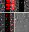
Figure 2. Exchange of membrane-RFP in SH-SY5Y cells through tunneling nanotubes (TNTs).
Some TNTs are visible with brightfield and membrane-RFP (blue arrows), whereas thinner TNTs are only seen with membrane-RFP (green arrows). (a) Formation of TNTs of variable thickness in SH-SY5Y cells. Membrane is exchanged with the other SH-SY5Y cell (yellow arrows), but gradually disappears throughout the recording. (b) Increased formation of TNTs in SH-SY5Y cell 1 during mitosis (blue and green arrows) with the substrate and the neighboring cells. No membrane exchange is observed during this mitosis (c) SEM image of SH-SY5Y cells connected by a TNT (orange arrow). (d) SEM image of a mitotic SH-SY5Y cell (pink arrow) connected by a TNT (orange arrow). Scale bars represent 10 μm.
Figure 3
Figure 3. Expression of α-synuclein (α-syn) in human pericytes stained with PDGFRβ and transfer through tunneling nanotubes.
(a) Distribution and localisation of α-syn precipitates and PDGFRβ, a pericyte-specific marker in human control and Parkinson’s disease middle temporal gyrus (MTG). (b) Confocal recording with orthogonal views of a pericyte cell with an internalized α-syn precipitate (yellow arrows) in the olfactory bulb (OFB). (c) Single confocal plane of a pericyte cell, adjacent to an α-syn precipitate in MTG. (d) In vivo TNT-like nanotubes (green arrow) in OFB labeled with Hoechst. (e) Transfer of α-syn-mCherry through tunneling nanotubes (TNTs) in pericytes in vitro. Two pericytes move away from each other thereby forming a TNT (yellow arrows) between cells 1 (traced in green) and 2 (traced in pink). After 406 min the TNT is no longer visible and two α-syn mCherry particles (blue arrows) remain in the non-transfected cell 2 until the end of the recording (486 minutes). (f) Exchange of membrane-RFP between pericytes before and during mitosis. Before mitosis two pericytes (cells 1 and 2) move away from each other, forming a TNT (blue arrow). Membrane (green arrow) is transferred to cell 2 and remains there throughout the recording. At 75 min cell 1 goes through mitosis and forms several connections (pink arrows) with the substrate and neighboring cells. Most of these connection/TNTs are not visible with brightfield. One TNT allows exchange of membrane (yellow arrow) with cell 3 and remains there throughout the recording (196 min). (g) Exchange of membrane-RFP between pericytes. After 51 min one of the pericytes (cell 2) moves away, forming a TNT (yellow arrow) between cells 1 and 2. The TNT breaks off and membrane-RFP (green arrow) is exchanged with cell 2. The membrane-RFP quickly dissipates and is no longer visible 20 min after transfer (at 72 min). Scale bars represent 10 μm (a–d) and 30 μm (e–g).
Figure 4
Figure 4. Exchange and internalization of membrane-RFP and F-actin-GFP through TNTs.
(a) Different TNTs are formed between cell 1 and 2 (colored arrows indicate individual TNTs). These TNTs allow transfer of F-actin-GFP surrounded by membrane-RFP to the accepting cell. The particles are exchanged at different time points and remain visible until the end of the recording (865 min). (b) Subsequent confocal recording with orthogonal views of the accepting cell 2 shows internalized actin-GFP. Membrane is stained with WGA (blue). Colored arrows show internalized particles (arrow colors correspond to colors used in F). Scale bars represent 30 μm (D, E) or 10 μm (A–C, F–G).
Figure 5
Figure 5. Boxplot representing lengths of TNTs as measured with brightfield (t; grey boxplots) or with membrane-RFP (r; red boxplots) in four SH-SY5Y cells (S1-S4) and four pericytes (P1-P4).
The table below the boxplot shows the number of actual observed membrane-RFP TNT-mediated transfers between these cells during the same period.
Figure 6
Figure 6. Characterization of tunneling nanotube (TNT) properties in SH-SY5Y cells.
(a) Transfer occurred through TNTs (yellow arrows) within 30 sec of dye filling, and hydrazide-488 gradually increased in cells 2, 3, and 4. At 12 min, prior to filling cell 2 with hydrazide-594 a small amount of bleed through is present in the red channel (blue arrow). Dye filling of cell 2 with hydrazide-594 began at 22 min, and by 31 min, the TNT connecting cells 1 and 2 filled up with hydrazide-594 (pink arrow). (b) Electrical connectivity of SH-SY5Y cells 1 and 2 (from figure A) connected through a TNT 45 min after the dye filling of cell 1 began. (c) Closed TNT: no transfer of hydrazide-488 through the TNT after 10 min of dye filling cell 1. Yellow arrows indicate TNTs. (d) Open TNT: cell 1 was simultaneously filled with dextran (10 kDa)-488 and hydrazide-594. After 6 min both dextran (10 kDa)-488 and hydrazide-594 had transferred through the TNT.

References

    1. Feng L. R., Federoff H. J., Vicini S. & Maguire‐Zeiss K. A. α‐Synuclein mediates alterations in membrane conductance: a potential role for α‐synuclein oligomers in cell vulnerability. Eur. J. Neurosci. 32, 10–17 (2010). - PMC - PubMed
    1. Angot E. et al.. Alpha-synuclein cell-to-cell transfer and seeding in grafted dopaminergic neurons in vivo. PLoS One 7, e39465–e39465 (2012). - PMC - PubMed
    1. Adler C. H. & Beach T. G. Neuropathological basis of nonmotor manifestations of Parkinson’s disease. Mov. Disord. (2016). - PMC - PubMed
    1. Li J.-Y. et al.. Lewy bodies in grafted neurons in subjects with Parkinson’s disease suggest host-to-graft disease propagation. Nat Med 14, 501–503 (2008). - PubMed
    1. Kordower J. H., Chu Y., Hauser R. A., Freeman T. B. & Olanow C. W. Lewy body-like pathology in long-term embryonic nigral transplants in Parkinson’s disease. Nat Med 14, 504–506 (2008). - PubMed

Publication types

MeSH terms

Substances