. 2017 Feb 23:7:43342.
doi: 10.1038/srep43342.

TubZ filament assembly dynamics requires the flexible C-terminal tail

Affiliations

TubZ filament assembly dynamics requires the flexible C-terminal tail

Maria E Fuentes-Pérez et al. Sci Rep. .

Abstract

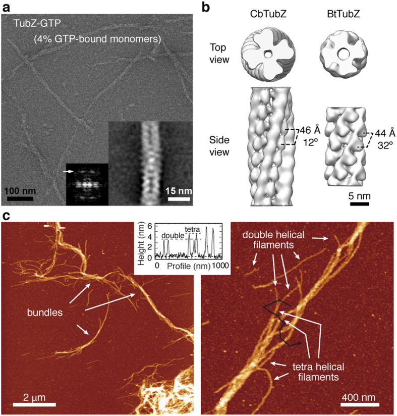
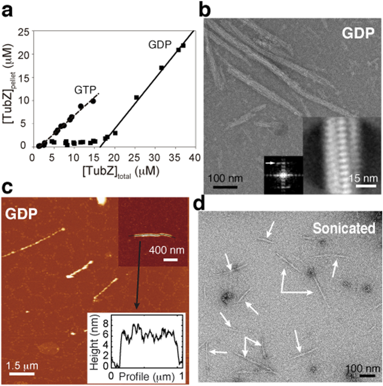
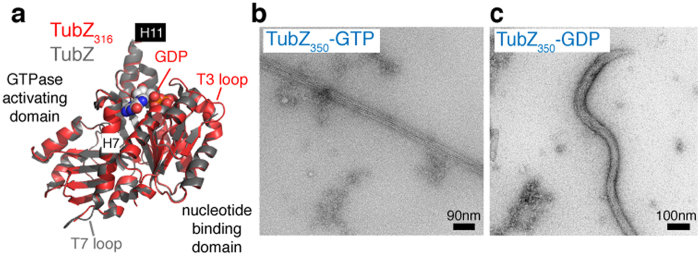
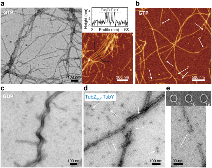
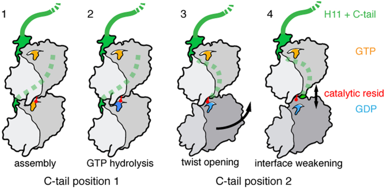
Cytomotive filaments are essential for the spatial organization in cells, showing a dynamic behavior based on nucleotide hydrolysis. TubZ is a tubulin-like protein that functions in extrachromosomal DNA movement within bacteria. TubZ filaments grow in a helical fashion following treadmilling or dynamic instability, although the underlying mechanism is unclear. We have unraveled the molecular basis for filament assembly and dynamics combining electron and atomic force microscopy and biochemical analyses. Our findings suggest that GTP caps retain the filament helical structure and hydrolysis triggers filament stiffening upon disassembly. We show that the TubZ C-terminal tail is an unstructured domain that fulfills multiple functions contributing to the filament helical arrangement, the polymer remodeling into tubulin-like rings and the full disassembly process. This C-terminal tail displays the binding site for partner proteins and we report how it modulates the interaction of the regulator protein TubY.


Conflict of interest statement

The authors declare no competing financial interests.

Figures

Figure 1
Figure 1. Characterization of TubZ filaments.
(a) Negative stain EM showing wild type CbTubZ filaments. Inset corresponds to the averaged filament and its Fourier transform. Arrow indicates a 4.8-nm longitudinal spacing between molecules. (b) Top and side views of our CbTubZ low-resolution filament versus BtTubZ filament filtered at a similar low-resolution level. Both filaments are 4-stranded but show different longitudinal distances between monomers and azimuthal angles. (c) AFM images of CbTubZT100A filaments, showing 2 – and 4-stranded filaments and bundles. The black line in the second image corresponds to the height profile shown in the graph. Color scale from dark to bright corresponds to 0–11 nm.
Figure 2
Figure 2. Effect of GTP hydrolysis on filament structure.
(a) Critical concentration measurements of CbTubZ in buffer containing Ficoll (200 g/L) and GTP/Mg2+ or GDP/Mg2+ (b). Negative stain EM showing CbTubZ filaments polymerized in the presence of GDP/Mg2+. Inset shows averaged filaments and the corresponding Fourier transform. Arrow indicates 4.8-nm longitudinal spacing between molecules. (c) AFM images of CbTubZ filaments polymerized in the presence of GDP/Mg2+. The black line corresponds to the height profile shown in the graph. Color scale from dark to bright correspond to 0–11 nm. (d) Negative stain EM of CbTubZ filaments assembled with GTP/Mg2+ and sonicated for 10 min, showing the stiffening effect.
Figure 3
Figure 3. Effect of C-tail on TubZ assembly.
(a) Comparison of full length and CbTubZ316 structures, highlighting the main domain arrangements and the most important secondary structure elements. TubZ316 displays GDP at the nucleotide-binding pocket, an ordered T3 loop and a disordered T7 loop (which is ordered in the full-length protein). In both structures the C-terminal helix H11 is in the same position, but neither contain an ordered C-tail. (b,c) Negative stain EM of CbTubZ350 stiff filament assembled in the presence of GTP/Mg2+ (b) and flexible filament polymerized with GDP/Mg2+ (c).
Figure 4
Figure 4. TubY - TubZ interaction.
(a) Negative stain EM (left) and AFM (right) of CbTubZ-TubY filaments. The height profile shown in the graph was taken in the direction of the black arrow. (b) AFM images of CbTubZE200A-TubY filaments. White arrows indicate thinner extensions at the ends of some filaments. Color scale from dark to bright is 0–11 nm in AFM images. (ce) EM images of TubY binding to GDP-bound CbTubZ filaments (c), TubZ350 filaments and rings formed in the presence of TubY (d) and detail of ring formation upon disassembly from filaments tips (e, bottom). (e) Different types of averaged structures obtained after ring processing, including the number of monomers in each ring (top).
Figure 5
Figure 5. Model of TubZ filament dynamics.
For simplicity, we show only two consecutive molecules in the filament: (1) GTP-bound state induces the assembly, whereas the C-tail (green and dashed line when behind a protein subunit) establishes specific contacts with upper subunits leading to the generation of a twist and allowing the formation of the canonical longitudinal interface; (2) the catalytic residue (red) is correctly positioned and reaches GTP (yellow), which is hydrolyzed into GDP (blue); (3) the enzymatic reaction induces changes in the bottom molecule, changing the C-tail interacting surface with the upper subunits and opening the twist between both molecules; (4) the GDP-bound state results in weakening of the longitudinal interface, likely due to loss of contacts. C-tail truncated proteins do not establish the specific contacts with upper subunits within the filament, blocking the formation of the canonical longitudinal interface. Accordingly, GTP assembly leads to state 3 filaments, while GDP-bound polymers are in state 4.

Similar articles

Cited by

References

    1. Nogales E., Downing K. H., Amos L. A. & Lowe J. Tubulin and FtsZ form a distinct family of GTPases. Nat Struct Biol 5, 451–458 (1998). - PubMed
    1. Oliva M. A. & Andreu J. M. In eLS Citable Reviews doi: 10.1002//9780470015902.a0025586 (2014). - DOI
    1. Larsen R. A. et al.. Treadmilling of a prokaryotic tubulin-like protein, TubZ, required for plasmid stability in Bacillus thuringiensis. Genes Dev 21, 1340–1352 (2007). - PMC - PubMed
    1. Baxter J. C. & Funnell B. E. Plasmid Partition Mechanisms. Microbiol Spectr 2, doi: 10.1128/microbiolspec.PLAS-0023-2014 (2014). - DOI - PubMed
    1. Oliva M. A., Martin-Galiano A. J., Sakaguchi Y. & Andreu J. M. Tubulin homolog TubZ in a phage-encoded partition system. Proc Natl Acad Sci USA 109, 7711–7716 (2012). - PMC - PubMed

Publication types