Review
. 2017 Feb 20;9(2):159.
doi: 10.3390/nu9020159.

Role of Vitamin A/Retinoic Acid in Regulation of Embryonic and Adult Hematopoiesis

Affiliations
Review

Role of Vitamin A/Retinoic Acid in Regulation of Embryonic and Adult Hematopoiesis

Ana Cañete et al. Nutrients. .

Abstract

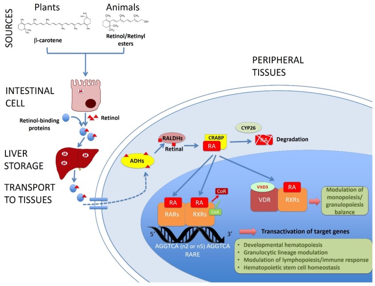
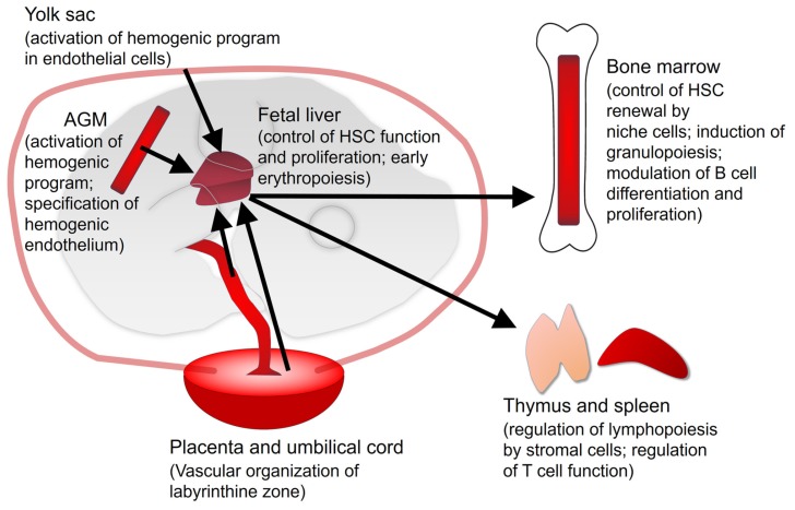
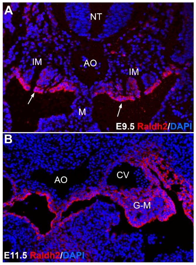
Vitamin A is an essential micronutrient throughout life. Its physiologically active metabolite retinoic acid (RA), acting through nuclear retinoic acid receptors (RARs), is a potent regulator of patterning during embryonic development, as well as being necessary for adult tissue homeostasis. Vitamin A deficiency during pregnancy increases risk of maternal night blindness and anemia and may be a cause of congenital malformations. Childhood Vitamin A deficiency can cause xerophthalmia, lower resistance to infection and increased risk of mortality. RA signaling appears to be essential for expression of genes involved in developmental hematopoiesis, regulating the endothelial/blood cells balance in the yolk sac, promoting the hemogenic program in the aorta-gonad-mesonephros area and stimulating eryrthropoiesis in fetal liver by activating the expression of erythropoietin. In adults, RA signaling regulates differentiation of granulocytes and enhances erythropoiesis. Vitamin A may facilitate iron absorption and metabolism to prevent anemia and plays a key role in mucosal immune responses, modulating the function of regulatory T cells. Furthermore, defective RA/RARα signaling is involved in the pathogenesis of acute promyelocytic leukemia due to a failure in differentiation of promyelocytes. This review focuses on the different roles played by vitamin A/RA signaling in physiological and pathological mouse hematopoiesis duddurring both, embryonic and adult life, and the consequences of vitamin A deficiency for the blood system.

Keywords: embryos; hematopoiesis; leukemia; retinoic acid; vitamin A; vitamin A deficiency.


Conflict of interest statement

The authors declare no conflict of interest.

Figures

Figure 1
Figure 1
Role of vitamin A/retinol in adult hematopoiesis. This picture shows the main molecular pathways leading from the vitamin A sources to the target genes of the retinoic acid (RA, the active form of vitamin A) related with hematopoiesis in the tissues. Retinol or provitamin A is ingested and absorbed through the intestine, transported by retinol-binding proteins and stored in the liver. Retinol is transformed by the cells into RA by alcohol and aldehyde dehydrogenases (ADHs and RALDHs respectively). RA is transported by cellular retinoic acid binding proteins (CRABP) and it can be degraded by CYP26 or translocated to the nucleus, where it binds and activates nuclear retinoid acid receptors (RARs and RXRs), displacing co-repressors and recruiting coactivators of the transcription of target genes. In this way, RA regulates the developmental hematopoiesis, modulates lympho and granulopoiesis and contributes to the homeostasis of the hematopoietic stem cells. Vitamin D receptor (VDR) can also dimerize with RXRs and modulate the immune response.
Figure 2
Figure 2
Hematopoietic sites in the embryo (left) and adult mouse (right). The main functions played by vitamin A/RA signaling are described between parentheses. Black arrows represent the main routes of migration of progenitors between the hematopoietic organs and tissues. Exchange of progenitors between embryonic organs is also possible. It is unknown if RA signaling plays an intrinsic role on placental hematopoietic stem cells (HSC). AGM: Aorta-Gonad-Mesonephros.
Figure 3
Figure 3
Vitamin A in developmental hematopoiesis. The picture shows immunolocalization of the enzyme RALDH2, catalyzing a key step in the generation of retinoic acid from retinol in the aorta-gonad-mesonephros. RALDH2 is expressed in the coelomic epithelium of the intermediate mesoderm (IM) (arrows in A) by the stage E9.5 (A) and later (B) in mesenchymal cells of area, where the definitive population of hematopoietic stem cells is generated, between the aorta (AO), the cardinal veins (CV) and the gonadal/mesonephric mesoderm (G-M). This embryonic RA signaling is essential for the emergence of the definitive blood progenitors, as described in the text. M: mesentery; NT: neural tube.

References

    1. Semba R.D. The discovery of the vitamins. Int. J. Vitam. Nutr. Res. 2012;823:10–15. doi: 10.1159/000343124. - DOI - PubMed
    1. Adamo S., De Luca L.M., Akalovsky I., Bhat P.V. Retinoid-induced adhesion in cultured, transformed mouse fibroblasts. J. Natl. Cancer Inst. 1979;62:1473–1478. - PubMed
    1. Kato S., De Luca L.M. Retinoic acid modulates attachment of mouse fibroblasts to laminin substrates. Exp. Cell Res. 1987;173:450–462. doi: 10.1016/0014-4827(87)90285-0. - DOI - PubMed
    1. De Luca L.M., Adamo S., Kato S. Retinoids and cell adhesion. Methods Enzymol. 1990;190:81–91. - PubMed
    1. Gerster H. Vitamin A-functions, dietary requirements and safety in humans. Int. J. Vitam. Nutr. Res. 1997;67:71–90. - PubMed

MeSH terms