Review
. 2017 Feb 20;6(2):22.
doi: 10.3390/jcm6020022.

CVD and Oxidative Stress

Affiliations
Review

CVD and Oxidative Stress

Karla Cervantes Gracia et al. J Clin Med. .

Abstract

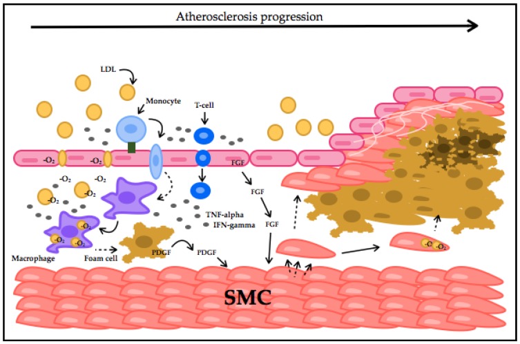
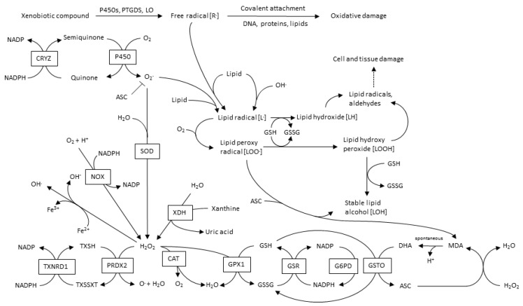
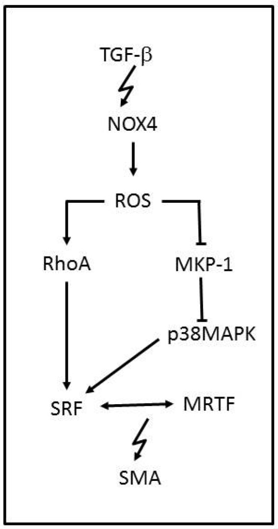
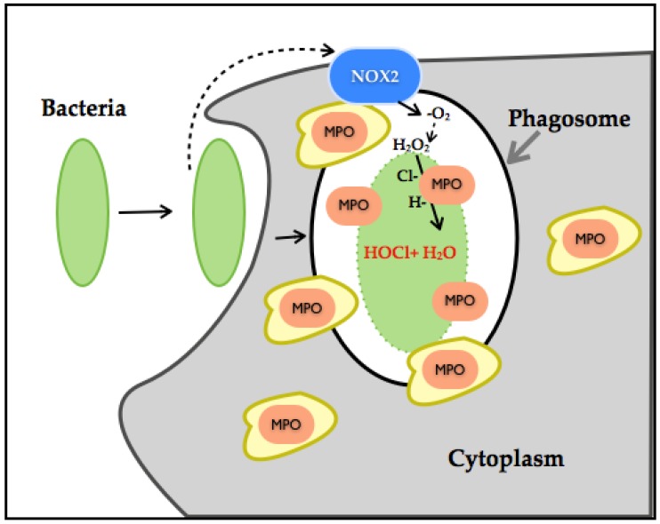
Nowadays, it is known that oxidative stress plays at least two roles within the cell, the generation of cellular damage and the involvement in several signaling pathways in its balanced normal state. So far, a substantial amount of time and effort has been expended in the search for a clear link between cardiovascular disease (CVD) and the effects of oxidative stress. Here, we present an overview of the different sources and types of reactive oxygen species in CVD, highlight the relationship between CVD and oxidative stress and discuss the most prominent molecules that play an important role in CVD pathophysiology. Details are given regarding common pharmacological treatments used for cardiovascular distress and how some of them are acting upon ROS-related pathways and molecules. Novel therapies, recently proposed ROS biomarkers, as well as future challenges in the field are addressed. It is apparent that the search for a better understanding of how ROS are contributing to the pathophysiology of CVD is far from over, and new approaches and more suitable biomarkers are needed for the latter to be accomplished.

Keywords: CVD; cardiovascular disease; oxidative stress; reactive oxygen species.


Conflict of interest statement

The authors declare no conflict of interest.

Figures

Figure 1
Figure 1
Atherosclerotic plaque formation process. LDLs at a high concentration inhibit endothelial cells’ (EC) endocytosis capacity, migrate and accumulate in the intima. LDLs get oxidized and induce VCAM (green square) expression in EC. Monocytes are recruited into the intima by VCAM interaction. Monocytes transform into macrophages, which take-up oxLDLs, forming foam cells. Macrophages and EC secrete chemokines and recruit T-cells. T-cells produce TNF-alpha and IFN-gamma, amplifying inflammation. Fibroblast growth factor (FGF) and platelet-derived growth factor (PDGF) stimulate smooth-muscle cells (SMC) migration and proliferation. SMCs can also accumulate lipids, migrate and proliferate. A lipid core is generated with necrotic foam cells surrounded by SMCs and a collagen fibrous cap, resulting in thrombus formation.
Figure 2
Figure 2
Metabolic pathways in ROS production and metabolism.
Figure 3
Figure 3
NADPH oxidase-modulated pathway in vascular smooth muscle cell differentiation. The TGF-β-mediated induction of NOX4 causes an inhibition of MKP-1 and activation of RhoA by ROS. Under physiological conditions, MKP-1 inhibits p38MAPK, thereby preventing the RhoA-mediated release and association of transcription elements SRF, which can be activated by p38MAPK, and MRTF, which drive SMA gene expression.
Figure 4
Figure 4
Illustration of neutrophil NOX-HOCl production. Neutrophils have phagosomes and granules in its cytoplasm. Granules contain myeloperoxidase (MPO) (orange). The NOX2 complex (blue) gets assembled once the neutrophils initiate bacterial endocytosis. NOX2 produces H2O2 by dismutation, and MPO reacts with H2O2 and chloride ions, thereby generating HOCl and water, leading to neutrophil bactericidal effects.
Figure 5
Figure 5
Molecular pathways associated with CVD etiology. Contributing CVD factors, such as diabetes, cause an imbalance of essential physiological pathways by impairing normal glycolysis (GL), which feeds into the tricarboxylic acid pathway (TCA) that is directly linked to the electron transport chain (ETC). Fatty acids, which can also contribute and feed into the TCA cycle through fatty acid oxidation (FAO), are also implicated in CVD manifestation. Multiple enzymes involved in this scheme have been shown to either modulate or directly generate ROS.

Similar articles

Cited by

References

    1. Sies H. Oxidative stress: A concept in redox biology and medicine. Redox Biol. 2015;4:180–183. doi: 10.1016/j.redox.2015.01.002. - DOI - PMC - PubMed
    1. Jones D.P. Redefining oxidative stress. Antioxid. Redox Signal. 2006;8:1865–1879. doi: 10.1089/ars.2006.8.1865. - DOI - PubMed
    1. Mittal C.K., Murad F. Activation of guanylate cyclase by superoxide dismutase and hydroxyl radical: A physiological regulator of guanosine 3′,5′-monophosphate formation. Proc. Natl. Acad. Sci. USA. 1977;74:4360–4364. doi: 10.1073/pnas.74.10.4360. - DOI - PMC - PubMed
    1. Olguin-Albuerne M., Moran J. Ros produced by NOX2 control in vitro development of cerebellar granule neurons development. ASN Neuro. 2015 doi: 10.1177/1759091415578712. - DOI - PMC - PubMed
    1. Mandal D., Fu P., Levine A.D. REDOX regulation of IL-13 signaling in intestinal epithelial cells: Usage of alternate pathways mediates distinct gene expression patterns. Cell. Signal. 2010;22:1485–1494. doi: 10.1016/j.cellsig.2010.05.017. - DOI - PMC - PubMed

LinkOut - more resources