

The Adverse Effects of Triptolide on the Reproductive System of Caenorhabditis elegans: Oogenesis Impairment and Decreased Oocyte Quality
- PMID: 28230788
- PMCID: PMC5343997
- DOI: 10.3390/ijms18020464
The Adverse Effects of Triptolide on the Reproductive System of Caenorhabditis elegans: Oogenesis Impairment and Decreased Oocyte Quality
Abstract
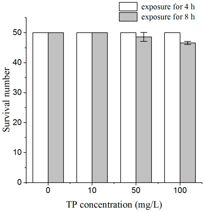
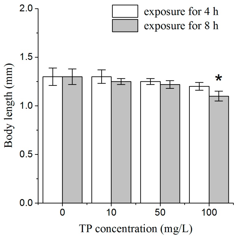
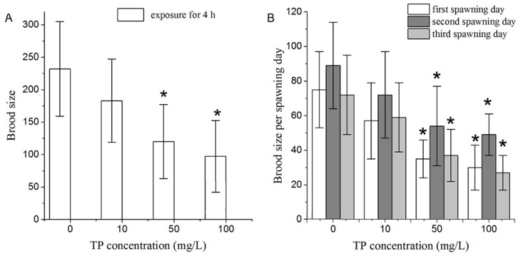
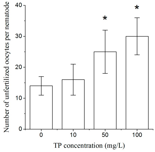
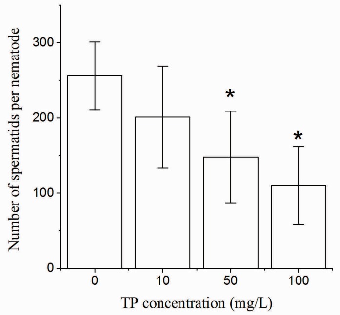
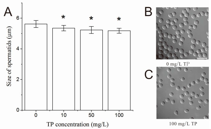
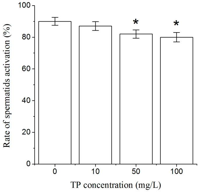
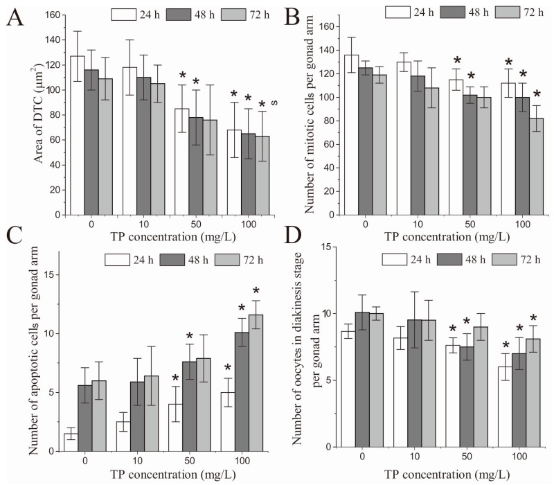
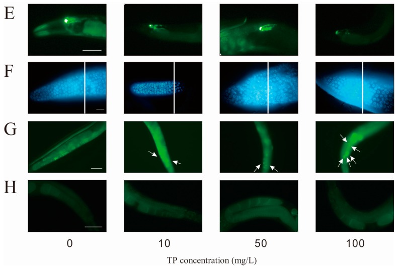
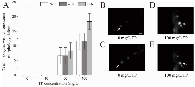
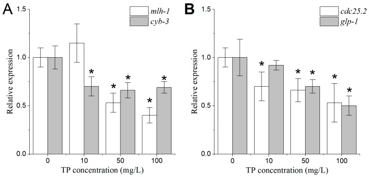
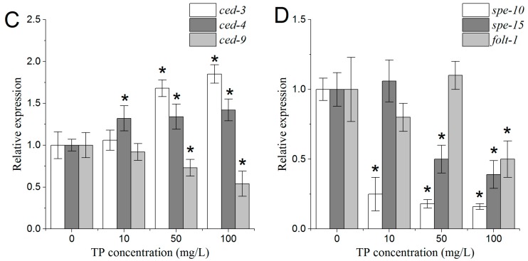
Previous studies have revealed that Triptolide damages female reproductive capacity, but the mechanism is unclear. In this study, we used Caenorhabditis elegans to investigate the effects of Triptolide on the germline and explore its possible mechanisms. Our data show that exposure for 4 h to 50 and 100 mg/L Triptolide reduced C. elegans fertility, led to depletion and inactivation of spermatids with the changes in the expression levels of related genes, and increased the number of unfertilized oocytes through damaging chromosomes and DNA damage repair mechanisms. After 24 and 48 h of the 4 h exposure to 50 and 100 mg/L Triptolide, we observed shrink in distal tip cells, an increase in the number of apoptotic cells, a decrease in the number of mitotic germ cells and oocytes in diakinesis stage, and chromatin aggregates in -1 oocytes. Moreover, expression patterns of the genes associated with mitotic germ cell proliferation, apoptosis, and oocyte quality were altered after Triptolide exposure. Therefore, Triptolide may damage fertility of nematodes by hampering the development of oocytes at different developmental stages. Alterations in the expression patterns of genes involved in oocyte development may explain the corresponding changes in oocyte development in nematodes exposed to Triptolide.
Keywords: Caenorhabditis elegans; Triptolide; apoptosis; mitosis; oogenesis.
Conflict of interest statement
The authors declare no conflict of interest.
Figures
Similar articles
-
Chlorpyrifos exposure reduces reproductive capacity owing to a damaging effect on gametogenesis in the nematode Caenorhabditis elegans.J Appl Toxicol. 2012 Jul;32(7):527-35. doi: 10.1002/jat.1783. Epub 2011 Dec 19. J Appl Toxicol. 2012. PMID: 22180373
-
Di (2-ethylhexyl) phthalate-induced reproductive toxicity involved in dna damage-dependent oocyte apoptosis and oxidative stress in Caenorhabditis elegans.Ecotoxicol Environ Saf. 2018 Nov 15;163:298-306. doi: 10.1016/j.ecoenv.2018.07.066. Epub 2018 Jul 26. Ecotoxicol Environ Saf. 2018. PMID: 30056344
-
Supplementation with Triptolide Increases Resistance to Environmental Stressors and Lifespan in C. elegans.J Food Sci. 2017 Jun;82(6):1484-1490. doi: 10.1111/1750-3841.13720. Epub 2017 May 4. J Food Sci. 2017. PMID: 28471052
-
[Progress in research on triptolide].Zhongguo Zhong Yao Za Zhi. 2005 Feb;30(3):170-4. Zhongguo Zhong Yao Za Zhi. 2005. PMID: 15719629 Review. Chinese.
-
Triptolide, A Potential Autophagy Modulator.Chin J Integr Med. 2019 Mar;25(3):233-240. doi: 10.1007/s11655-018-2847-z. Epub 2018 Sep 4. Chin J Integr Med. 2019. PMID: 30178091 Review.
Cited by
-
Critical Role of Hepatic Cyp450s in the Testis-Specific Toxicity of (5R)-5-Hydroxytriptolide in C57BL/6 Mice.Front Pharmacol. 2017 Nov 21;8:832. doi: 10.3389/fphar.2017.00832. eCollection 2017. Front Pharmacol. 2017. PMID: 29209210 Free PMC article.
-
Exploiting DNA repair defects in triple negative breast cancer to improve cell killing.Ther Adv Med Oncol. 2020 Sep 18;12:1758835920958354. doi: 10.1177/1758835920958354. eCollection 2020. Ther Adv Med Oncol. 2020. PMID: 32994807 Free PMC article.
-
Triptolide potentiates the cytoskeleton-stabilizing activity of cyclosporine A in glomerular podocytes via a GSK3β dependent mechanism.Am J Transl Res. 2020 Mar 15;12(3):800-812. eCollection 2020. Am J Transl Res. 2020. PMID: 32269713 Free PMC article.
-
The Anti-Inflammatory and Immunomodulatory Activities of Natural Products to Control Autoimmune Inflammation.Int J Mol Sci. 2022 Dec 21;24(1):95. doi: 10.3390/ijms24010095. Int J Mol Sci. 2022. PMID: 36613560 Free PMC article. Review.
-
Andrographolide as a Therapeutic Agent Against Breast and Ovarian Cancers.Open Life Sci. 2019 Dec 4;14:462-469. doi: 10.1515/biol-2019-0052. eCollection 2019 Jan. Open Life Sci. 2019. PMID: 33817182 Free PMC article.
References
-
- Li Z.K., Wang C.Z., Qian G.S., Lin K.X., Liu G. Regulating effect of Triptolide on expression of IL-5, IL-3 and GM-CSF receptors mRNA in BALF eosinophils of asthmatic guinea pigs. Immunol. J. 2002;18:102–106.
-
- Yang S., Chen J., Gu Z., Xu X.M., Wang L., Pei X.F., Yang J., Underhill C.B., Zhang L. Triptolide inhibits the growth and metastasis of solid tumors. Mol. Cancer Ther. 2003;2:65–72. - PubMed
MeSH terms
Substances
LinkOut - more resources
Full Text Sources
Other Literature Sources
