. 2017 Feb 21;14(2):213.
doi: 10.3390/ijerph14020213.

Characterization of Carbonic Anhydrase 9 in the Alimentary Canal of Aedes aegypti and Its Relationship to Homologous Mosquito Carbonic Anhydrases

Affiliations

Characterization of Carbonic Anhydrase 9 in the Alimentary Canal of Aedes aegypti and Its Relationship to Homologous Mosquito Carbonic Anhydrases

Daniel P Dixon et al. Int J Environ Res Public Health. .

Abstract

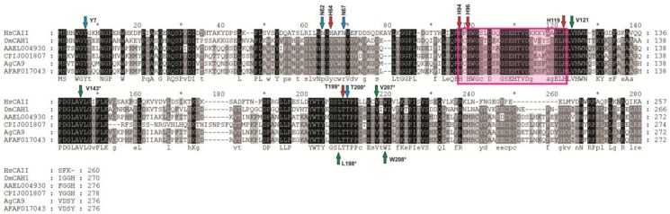
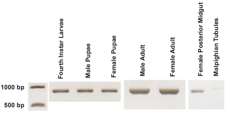
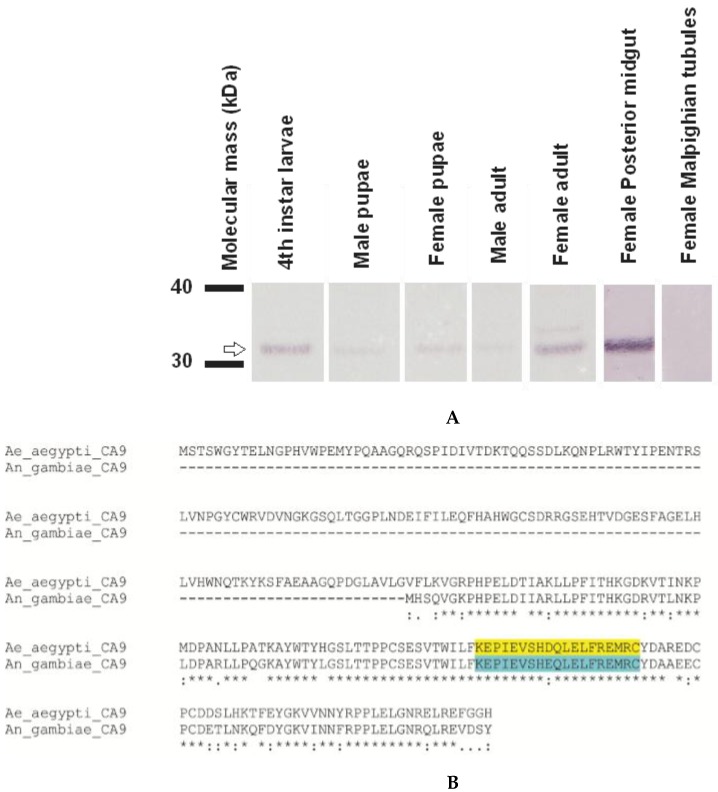
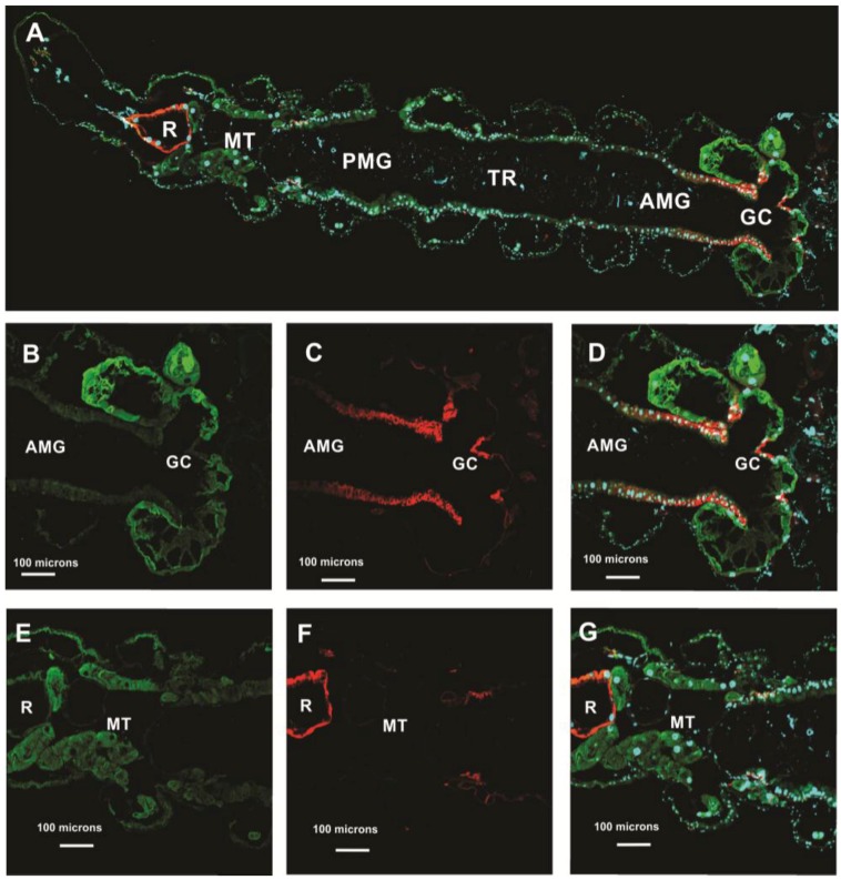
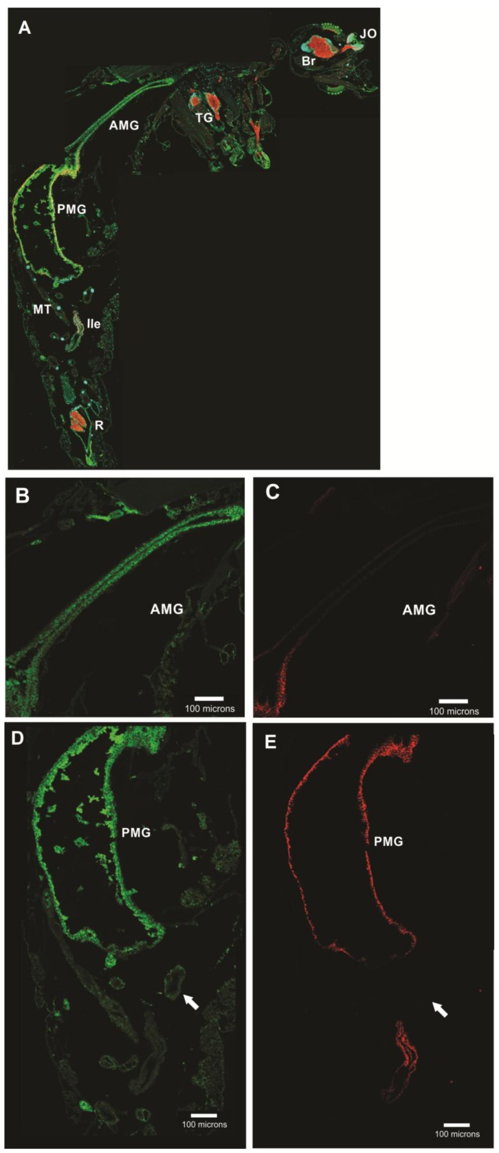
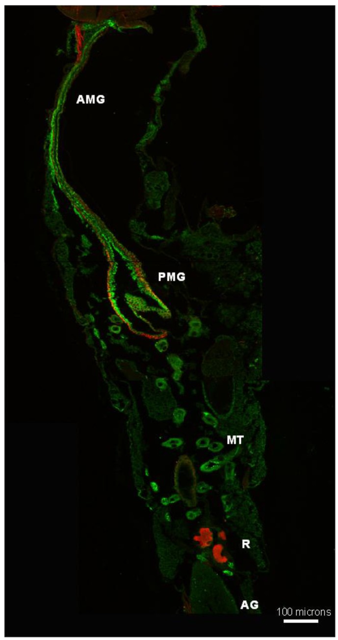
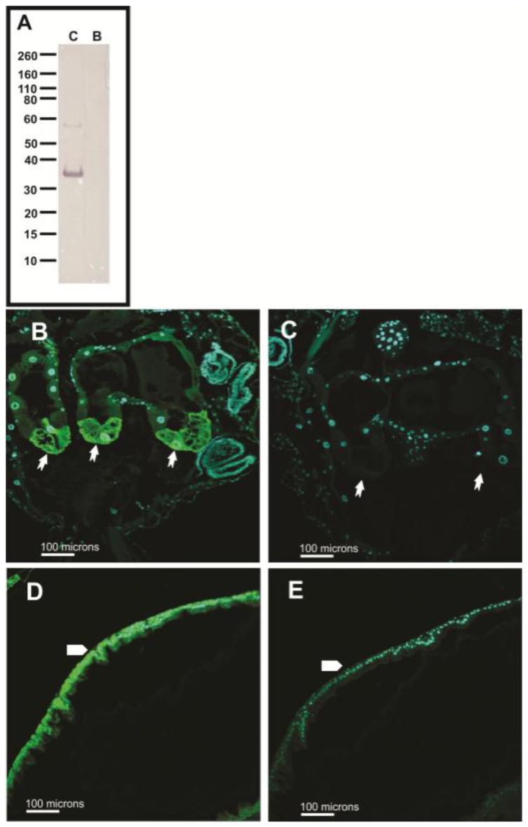
In the mosquito midgut, luminal pH regulation and cellular ion transport processes are important for the digestion of food and maintenance of cellular homeostasis. pH regulation in the mosquito gut is affected by the vectorial movement of the principal ions including bicarbonate/carbonate and protons. As in all metazoans, mosquitoes employ the product of aerobic metabolism carbon dioxide in its bicarbonate/carbonate form as one of the major buffers of cellular and extracellular pH. The conversion of metabolic carbon dioxide to bicarbonate/carbonate is accomplished by a family of enzymes encoded by the carbonic anhydrase gene family. This study characterizes Aedes aegypti carbonic anhydrases using bioinformatic, molecular, and immunohistochemical methods. Our analyses show that there are fourteen Aedes aegypti carbonic anhydrase genes, two of which are expressed as splice variants. The carbonic anhydrases were classified as either integral membrane, peripheral membrane, mitochondrial, secreted, or soluble cytoplasmic proteins. Using polymerase chain reaction and Western blotting, one of the carbonic anhydrases, Aedes aegypti carbonic anhydrase 9, was analyzed and found in each life stage, male/female pupae, male/female adults, and in the female posterior midgut. Next, carbonic anhydrase 9 was analyzed in larvae and adults using confocal microscopy and was detected in the midgut regions. According to our analyses, carbonic anhydrase 9 is a soluble cytoplasmic enzyme found in the alimentary canal of larvae and adults and is expressed throughout the life cycle of the mosquito. Based on previous physiological analyses of adults and larvae, it appears AeCA9 is one of the major carbonic anhydrases involved in producing bicarbonate/carbonate which is involved in pH regulation and ion transport processes in the alimentary canal. Detailed understanding of the molecular bases of ion homeostasis in mosquitoes will provide targets for novel mosquito control strategies into the new millennium.

Keywords: alkaline; carbonic anhydrase (CA); data mining; immunohistochemistry; midgut; molecular phylogenetics; mosquito; pH.


Conflict of interest statement

The authors declare no conflict of interest.

Figures

Figure 1
Figure 1
Anatomy of the larval and adult mosquito gut. (A) The larval alimentary canal is comprised of the foregut and cardia (not shown), GC (1), AMG (2), PMG (3), MTs (4), ileum (5), and rectum (6) (adapted from [5]); (B) The adult alimentary canal is comprised of the AMG (2), PMG (3), MTs (4), ileum (5), rectum (6), and rectal papillae (7).
Figure 2
Figure 2
Transcriptomic analysis of CA in each life stage via RNA-seq [6]. The rows indicate each CA gene, while each column is a life stage analyzed from the [6] transcriptome. The color on the heat map represents the relative expression in Reads per Kilobase per Million Mapped Reads (RPKM) of each CA in each tissue. The more hot colors (reds and orange) represent more abundant expression, while cooler colors (blues and greens) represent less abundant expression. A legend to the right of the heat map provides a reference for the color code to RPKM levels.
Figure 3
Figure 3
Transcriptomic analysis of CA in the carcass and ovaries post blood meal via RNA-seq [6]. The rows indicate each CA gene, while each column is a blood meal time point in either the carcass or ovaries analyzed from the [6] transcriptome. The color on the heat map represents the relative expression in Reads per Kilobase per Million Mapped Reads (RPKM) of each CA in each tissue and time point. The more hot colors (reds and orange) represent more abundant expression, while cooler colors (blues and greens) represent less abundant expression. A legend to the right of the heat map provides a reference for the color code to RPKM levels.
Figure 4
Figure 4
Molecular phylogeny of Aedes aegypti CA isoforms. This maximum likelihood molecular phylogeny was generated using MEGA 6 and based on a MUSCLE alignment of Homo sapiens, Drosophila melanogaster, Aedes aegypti, Culex quinquefasciatus, Anopheles gambiae, and Anopheles farauti. Bootstrap values are represented by yellow circles (50%–75% bootstrap) and black squares (75%–100% bootstrap). Colored bubbles encircle branches of the phylogeny where CAs with similar cellular compartmentalization or structure were grouped together. A legend below the phylogeny shows which color pertains to which compartmentalization/structure. This phylogeny correlates well with the [4] phylogeny and now shows the relationship of Aedes aegypti CAs to Culex quinquefasciatus and Anopheles farauti CAs together with the other species. Finally, the tissue and cellular compartments within which each AeCA was expressed [6] is marked on the phylogeny to correlate RNA expression with the molecular phylogeny. The species color code is as follows: Homo sapiens (black), Drosophila melanogaster (blue), Aedes aegypti (red), Culex quinquefasciatus (green), Anopheles gambiae (orange), and Anopheles farauti (purple).
Figure 5
Figure 5
MUSCLE alignment of AeCA9 and its orthologues. AeCA9 (AAEL004930) was aligned with its orthologous sequences in Homo sapiens (HsCAII), Drosophila melanogaster (DmCAH1), Culex quinquefasciatus (CPIJ001807), Anopheles gambiae (AgCA9), and Anopheles farauti (AFAF017043). Homo sapiens CAII was used as a guide to map out the residues in Aedes aegypti that make up the active site and support active site structure (red arrows). Also, amino acids that stabilize the active site water molecules (blue arrows) and form the CO2 binding pocket (green arrows) were mapped onto the alignment. The residues are highlighted in a graded fashion using the following shades that represent amino acid percent identity: Black (100% identity), Dark gray (80% identity), Light gray (60% identity), no shade (no similarity). The pink box overlaying the alignment indicates where the Human CAII active site is relative to the insect CAs (Human His94 through His119).
Figure 6
Figure 6
PCR of AeCA9 in each life stage, female posterior midgut, and female Malpighian tubules. The transcript for AeCA9 is 831 bp, and the 1 KB DNA ladder (left) indicates the size of the PCR product. The PCR for each life stage and the posterior midgut was run for 30 cycles and is non-quantitative, while the PCR for the Malpighian tubules were run for 36 cycles and was also not quantitative. AeCA9 was detected in all the life stages analyzed, and AeCA9 transcript was expressed in the PMG and is barely detectable in the MTs.
Figure 7
Figure 7
Western blot of AeCA9 in each life stage, female posterior midgut, and female Malpighian tubules. (A) In this western blot, the same life stages, sexes, and tissues were analyzed as in Figure 7. 6 μg of total protein was loaded for the life stages (4th instar larvae, Male pupae, Female pupae, Male adult, Female adult) and 40 μg of total protein was loaded for the female posterior midugt and female Malpighian tubules. AeCA9 was detected in all the life stage and tissue samples analyzed except the MTs (top row). There is a higher molecular weight band in the female adult sample, but that could be due to over-development; (B) This is an alignment showing the antigenic sequence used to develop the antisera targeting AeCA9 in (A) and all subsequent immunohistochemical analyses. The antisera was generated in chicken to target Anopheles gambiae CA9, and it has suitable cross-reactivity with Aedes aegypti as shown. Take note that there is only 1 amino acid difference in 19 between the antigenic sites of AgCA9 and AeCA9.
Figure 8
Figure 8
CA9 is compartmentalized to the GC and MTs of Aedes aegypti larvae. This figure shows transverse sections of a 4th instar larvae. The sections were immunostained with antibodies targeting the following: AeCA9 (green), NaK-ATPase (red), and nuclei were counterstained with DAPI (cyan). (A) A montage of the entire alimentary canal which is comprised of the following regions: Gastric caeca (GC), Anterior midgut (AMG), transitional region (TR), posterior midgut (PMG), Malpighian tubules (MTs), and rectum (R). In BD, the GC is either immunostained with AeCA9 (B), NaK-ATPase (C), or a merge of AeCA9, NaK-ATPase, and DAPI (D). In B, AeCA9 is detected in all the cells of the GC except for the cells that make up the stem (arrowhead). AeCA9 is detected in the cap cells (arrow), and the signal for AeCA9 in the caecal lumen (*) is low. In EG, the MTs are either immunostained with AeCA9 (E), NaK-ATPase (F), or a merge of AeCA9, NaK-ATPase, and DAPI (G). AeCA9 could be detected in the distal MT (arrow with tail), but not as much in the proximal MT (flagged arrow). In both parts of the MTs, signal intensity for AeCA9 is low. There was no immunoreactivity of AeCA9 in the rectum, as was previously demonstrated by Smith et al. 2008 [40]. The scale bars in A–G represent 100 microns.
Figure 9
Figure 9
AeCA9 is compartmentalized to the female PMG and MTs. This figure shows transverse sections of a female adult. These sections were immunostained with antibodies targeting the following: AeCA9 (green), NaK-ATPase (red), and DAPI (cyan). (A) In the montage, the following organs can be seen: Johnston′s organ (JO), Brain (Br), thoracic ganglia (TG), anterior midgut (AMG), posterior midgut (PMG), Malpighian tubules (MT), ileum (Ile), and rectum (R); (B) AeCA9 is detectable in the cytoplasm throughout the length of the AMG; (C) NaK-ATPase is minimally expressed throughout the length of the AMG; (D) AeCA9 is detected in the PMG, while a faint signal for AeCA9 is detected in the MTs (arrow); (E) NaK-ATPase is detected on the basal membranes of the PMG epithelial cells and the ileum. The scale bars in AE represent 100 microns.
Figure 10
Figure 10
AeCA9 is compartmentalized to the alimentary canal of male Aedes aegypti. This image is a montage of the adult male alimentary canal, showing the anterior midgut (AMG), posterior midgut (PMG), Malpighian tubules (MT), Rectum (R), and accessory glands (AG). AeCA9 (green) and NaK-ATPase (red) are used to immunostain this section of a male mosquito, and AeCA9 signal was detected in the AMG, PMG, and MTs. NaK-ATPase was detected in the PMG, part of the AMG, and the rectal papillae.
Figure 11
Figure 11
Pre-absorption controls of AeCA9 antibody targeting larvae and adults. In this figure, the AeCA9 primary antibody was pre-absorbed with its associated peptide to determine its specificity in larvae and adults. (A) The protein used in the western blot is isolated PMG protein. Non-absorbed antiserum targeted lane C (control), while absorbed antiserum targeted lane B (blocked/absorbed). AeCA9 is around 32 kDa. AeCA9 was detected using the control antiserum, and the blocked antiserum did not detect AeCA9; (BE) In the sections, either larval GC (B & C) or the adult PMG (D & E) were targeted. The same antibody solution sets were used in the control (B & D) and blocked (C & E) tissue sections and western blots. In both the larval GC and adult PMG, AeCA9 signal was blocked after preabsorption with the peptide. In the larval GC, arrows indicate cap cells. In the adult PMG, the arrows represent the epithelial cells of the PMG. The green signal is for AeCA9 while the cyan signal is for DAPI.

Similar articles

Cited by

References

    1. Noriega F.G., Edgar K.A., Bechet R., Wells M.A. Midgut exopeptidase activities in Aedes aegypti are induced by blood feeding. J. Insect Physiol. 2002;48:205–212. doi: 10.1016/S0022-1910(01)00165-2. - DOI - PubMed
    1. Dadd R.H. Alkalinity within the midgut of mosquito larvae with alkaline-active digestive enzymes. J. Insect Physiol. 1975;21:1847–1853. doi: 10.1016/0022-1910(75)90252-8. - DOI - PubMed
    1. Del Pilar Corena M., VanEkeris L., Salazar M.I., Bowers D., Fiedler M.M., Silverman D., Tu C., Linser P.J. Carbonic anhydrase in the adult mosquito midgut. J. Exp. Biol. 2005;208:3263–3273. doi: 10.1242/jeb.01739. - DOI - PubMed
    1. Smith K.E., VanEkeris L.A., Linser P.J. Cloning and characterization of AgCA9, a novel alpha-carbonic anhydrase from Anopheles gambiae Giles sensu stricto (Diptera: Culicidae) larvae. J. Exp. Biol. 2007;210:3919–3930. doi: 10.1242/jeb.008342. - DOI - PubMed
    1. Linser P.J., Smith K.E., Seron T.J., Neira Oviedo M. Carbonic anhydrases and anion transport in mosquito midgut pH regulation. J. Exp. Biol. 2009;212:1662–1671. doi: 10.1242/jeb.028084. - DOI - PMC - PubMed

LinkOut - more resources