Review
. 2017 Feb 21;7(1):20.
doi: 10.3390/biom7010020.

m1A Post-Transcriptional Modification in tRNAs

Affiliations
Review

m1A Post-Transcriptional Modification in tRNAs

Stephanie Oerum et al. Biomolecules. .

Abstract

To date, about 90 post-transcriptional modifications have been reported in tRNA expanding their chemical and functional diversity. Methylation is the most frequent post-transcriptional tRNA modification that can occur on almost all nitrogen sites of the nucleobases, on the C5 atom of pyrimidines, on the C2 and C8 atoms of adenosine and, additionally, on the oxygen of the ribose 2'-OH. The methylation on the N1 atom of adenosine to form 1-methyladenosine (m1A) has been identified at nucleotide position 9, 14, 22, 57, and 58 in different tRNAs. In some cases, these modifications have been shown to increase tRNA structural stability and induce correct tRNA folding. This review provides an overview of the currently known m1A modifications, the different m1A modification sites, the biological role of each modification, and the enzyme responsible for each methylation in different species. The review further describes, in detail, two enzyme families responsible for formation of m1A at nucleotide position 9 and 58 in tRNA with a focus on the tRNA binding, m1A mechanism, protein domain organisation and overall structures.

Keywords: 1-methyladenosine; Trm10; Trm6–Trm61; TrmI; Trmt10C; m1A; tRNA, methylation.


Conflict of interest statement

The authors declare no conflict of interest.

Figures

Figure 1
Figure 1
m1A modifications in tRNAs. (A) Chemical structure of the m1A modification on the adenosine base; (B) The m1A modification shown on a tRNA at all sites where the modification occurs (in red). The domain in which the modification has been identified is indicated as A: archaea, E: eukaryotes, and B: bacteria. Some modifications are found in mitochondria which is indicated as M.
Figure 2
Figure 2
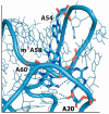
An overview of the ‘corner’ of the L-shaped initiator tRNA (tRNAi) from eukaryotes (PDB 1YFG). The nucleobases A20, A54, and A60 are highlighted along with the N1-modified A58 (m1A58). Hydrogen bonds are drawn as dotted lines.
Figure 3
Figure 3
Topology diagrams of the classical folds for (A) the SPOUT superfamily (two subtypes), and (B) the Rossmann-fold methyltransferase (RFM) superfamily. SAM: S-adenosyl-l-methionine.
Figure 4
Figure 4
Mechanism for N1-methylation on purine bases using the cofactor S-adenosyl-l-methionine (SAM) converting it to S-adenosyl-l-homocysteine (SAH). (A) Mechanism for m1G37 formation by TrmD (with aspartate as catalytic base) or Trm5 (with glutamate as catalytic base). Only the guanine base of tRNA is depicted for clarity. (B) Suggested mechanism for m1A based on studies on TrmI (m1A58) with aspartate as the catalytic base. (C) Alternative mechanism for m1A based on studies on TrmI (m1A58) where aspartate serves merely to position the cofactor and methylation site for reaction.
Figure 5
Figure 5
Structures of m1A58 tRNA MTases: (A) Trm6–Trm61 heterotetramer (PDB 5ERG); (B) Trm6–Trm61 heterotetramer bound to tRNALys3 (PDB 5CCB). The insert shows a superposition of free tRNALys3 (PDB 1FIR) and bound tRNALys3 (from PDB 5CCB). A58 and G19 are highlighted to emphasise the structural rearrangement of the tRNA upon binding; (C) TrmI homotetramer (PDB 2PWY).
Figure 6
Figure 6
Structural data of Trm10. (A) Full-length Trm10 from Sulfolobus acidocaldarius (PDB 5A7Y). Residues 182–202 are not modelled. (B) Docking from [84] of Escherichia coli initiator tRNA (PDB: 3CW5) onto Trm10 from S. acidocaldarius.
Figure 7
Figure 7
The electrostatic potential of the SPOUT domain for each Trm10 member for which a crystal structure is available. The SAM cofactor is shown as yellow sticks. The N1-methylation specificity is shown for each Trm10 member.

Similar articles

Cited by

References

    1. Phizicky E.M., Hopper A.K. tRNA biology charges to the front. Genes Dev. 2010;24:1832–1860. doi: 10.1101/gad.1956510. - DOI - PMC - PubMed
    1. Hopper A.K., Phizicky E.M. tRNA transfers to the limelight. Genes Dev. 2003;17:162–180. doi: 10.1101/gad.1049103. - DOI - PubMed
    1. Motorin Y., Helm M. tRNA stabilization by modified nucleotides. Biochemistry. 2010;49:4934–4944. doi: 10.1021/bi100408z. - DOI - PubMed
    1. Li H. Complexes of tRNA and maturation enzymes: Shaping up for translation. Curr. Opin. Struct. Biol. 2007;17:293–301. doi: 10.1016/j.sbi.2007.05.002. - DOI - PubMed
    1. Hopper A.K. Transfer RNA post-transcriptional processing, turnover, and subcellular dynamics in the yeast Saccharomyces cerevisiae. Genetics. 2013;194:43–67. doi: 10.1534/genetics.112.147470. - DOI - PMC - PubMed

Publication types

LinkOut - more resources