. 2017 Feb 23;8(2):e2626.
doi: 10.1038/cddis.2016.472.

Simvastatin-induced cell cycle arrest through inhibition of STAT3/SKP2 axis and activation of AMPK to promote p27 and p21 accumulation in hepatocellular carcinoma cells

Affiliations

Simvastatin-induced cell cycle arrest through inhibition of STAT3/SKP2 axis and activation of AMPK to promote p27 and p21 accumulation in hepatocellular carcinoma cells

Sin-Ting Wang et al. Cell Death Dis. .

Abstract

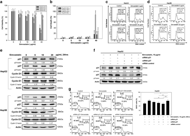
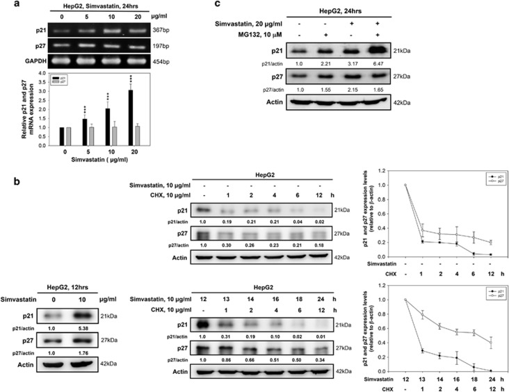
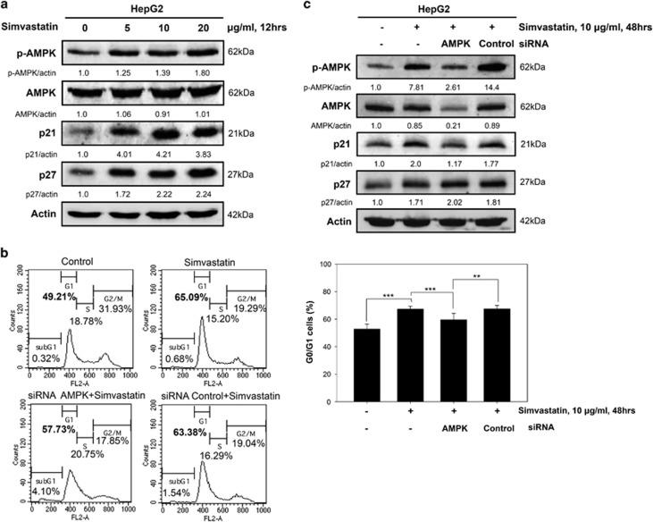
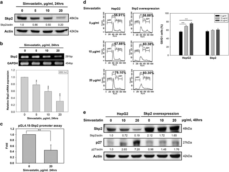
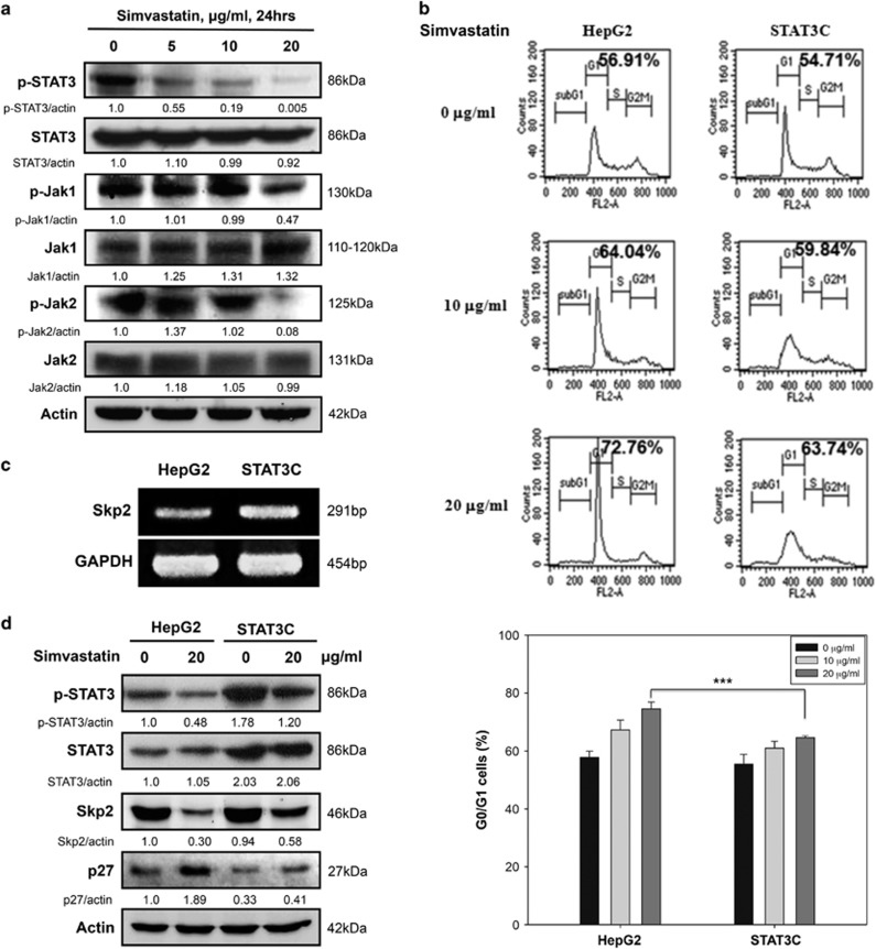
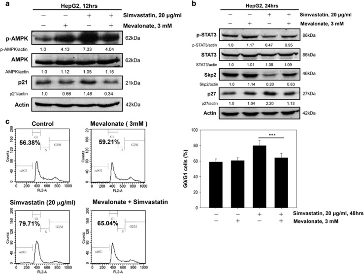
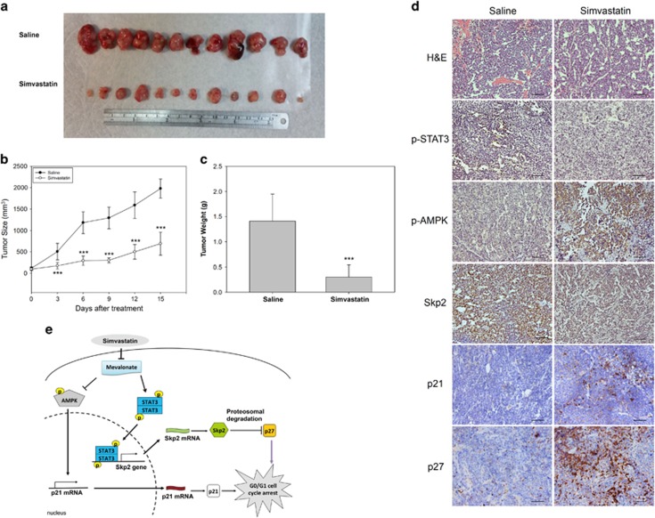
Hepatocellular carcinoma (HCC) is characterized by a poor prognosis and is one of the leading causes of cancer-related death worldwide. Simvastatin, an HMG-CoA reductase inhibitor, which decreases cholesterol synthesis by inhibiting mevalonate pathways and is widely used to treat cardiovascular diseases. Simvastatin exhibits anticancer effects against several malignancies. However, the molecular mechanisms underlying the anticancer effects of simvastatin on HCC are still not well understood. In this study, we demonstrated simvastatin-induced G0/G1 arrest by inducing p21 and p27 accumulation in HepG2 and Hep3B cells. Simvastatin also promoted AMP-activated protein kinase (AMPK) activation, which induced p21 upregulation by increasing its transcription. Consistent with this finding, we found genetic silencing of AMPK reduced p21 expression; however, AMPK silencing had no effect on p27 expression in HCC cells. Simvastatin decreased Skp2 expression at the transcriptional level, which resulted in p27 accumulation by preventing proteasomal degradation, an effect mediated by signal transducer and activator of transcription 3 (STAT3) inhibition. Constitutive STAT3 activation maintained high-level Skp2 expression and lower level p27 expression and significantly prevented G0/G1 arrest in simvastatin-treated HCC cells. Mevalonate decreased simvastatin-induced AMPK activation and rescued phospho-STAT3 and Skp2 expression in HCC cells, which resulted in the prevention of G0/G1 arrest through inhibition of p21 and p27 accumulation. Moreover, simvastatin significantly decreased tumor growth in HepG2 xenograft mice. Consistently, we found that simvastatin also increased p21 and p27 expression in tumor sections by reducing Skp2 expression and inducing AMPK activation and STAT3 suppression in the same tumor tissues. Taken together, these findings are demonstrative of the existence of a novel pathway in which simvastatin induces G0/G1 arrest by upregulating p21 and p27 by activating AMPK and inhibiting the STAT3-Skp2 axis, respectively. The results identify novel targets that explain the beneficial anticancer effects of simvastatin treatment on HCC in vitro and in vivo.


Conflict of interest statement

The authors declare no conflict of interest.

Figures

Figure 1
Figure 1
Simvastatin induces p21 and p27-dependent G0/G1 cell cycle arrest in HCC cell lines. Simvastatin suppressed cell growth in HCC cells. HepG2 and Hep3B cells were treated with various concentrations of simvastatin for 24 and 48 h. (a) Cell growth inhibition was measured by CCK-8 assay. (b) Cell death rates were determined by viable cell counting. These data are presented as percentages of vehicle-treated cells (*un-treated versus treated of HepG2 or Hep3B cells for 24 h or 48 h). (c and d) Simvastatin-induced HCC cell G0/G1 phase arrest. HepG2 and Hep3B cells were treated with simvastatin (0, 5, 10 or 20 μg/ml) for 48 h, and then cell cycle distributions were analyzed by PI staining and flow cytometry. (e) Simvastatin-induced G0/G1 phase-related protein expression in HCC cells. The cells were treated with simvastatin (0, 5, 10 or 20 μg/ml) for 24 h, and then the cell lysates were harvested for analysis of the expression of the cell cycle-related proteins p21, p27, cyclin D1, cyclin E1 and β-actin by immunoblotting. (f and g) Simvastatin-induced p21- and p27-dependent G0/G1 cell cycle arrest in HCC cells. HepG2 cells were transfected with p21, p27 or control siRNA for 24 h and then treated with 10 μg/ml simvastatin for 48 h. The cells were then harvested, and their DNA content and protein expression were analyzed by flow cytometry and immunoblotting using p21, p27 and β-actin antibodies. Data are expressed as the mean±S.E.M. of three independent experiments. Statistically significant differences between the un-treated and treated groups are indicated. *P<0.05, **P<0.01, ***P<0.001
Figure 2
Figure 2
Simvastatin-induced p21 and p27 protein upregulation was associated with transcriptional activation and protein degradation inhibition, respectively. (a) Simvastatin modulated p21 mRNA expression. HepG2 cells were treated with simvastatin (0, 5, 10 or 20 μg/ml) for 24 h, and p21, p27 and GAPDH mRNA expression levels were detected by RT-PCR and real-time PCR. (b) The effects of p21 and p27 protein stability in simvastatin-treated HCC cells. HepG2 cells were treated with 10 μg/ml CHX alone for 1, 2, 4, 6 or 12 h or 10 μg/ml simvastatin for 12 h. After 12 h, simvastatin-treated cells were co-treated with 10 μg/ml CHX for 1, 2, 4, 6 or 12 h. The cell lysates were harvested to detect p21, p27 and β-actin protein expression by immunoblotting. The intensity of each protein signal was determined by ImageJ software (downloaded from the NIH website (http://rsb.info.nih.gov/ij)). (c) Inhibition of proteasomal degradation promoted p21 accumulation, but not p27 accumulation, in simvastatin-treated HCC cells. HepG2 cells were treated with 20 μg/ml simvastatin with or without 10 μM MG132 for 24 h and then subjected to immunoblotting for the detection of p21, p27 and β-actin expression levels. Data are expressed as the mean±S.E.M. of three independent experiments. Statistically significant differences between the un-treated and treated groups are indicated. **P<0.01, ***P<0.001
Figure 3
Figure 3
Simvastatin-induced AMPK activation and p21 upregulation partially induced G0/G1 arrest in HepG2 cells. (a) Simvastatin-induced AMPK activation was associated with p21 and p27 upregulation. HepG2 cells were treated with simvastatin (0, 5, 10 or 20 μg/ml) for 12 h, and then immunoblotting was used to detect p-AMPK, AMPK, p21, p27 and β-actin protein expression levels. (b and c) Genetic silencing of AMPK reduced G0/G1 phase arrest and p21 expression in simvastatin-treated HCC cells. HepG2 cells were transfected with AMPK or control siRNA for 24 h and then treated with 10 μg/ml simvastatin for 48 h. (b) Cells were collected for analysis of their DNA content by flow cytometry. (c) Cell lysates were harvested to detect protein expression levels by immunoblotting using p-AMPK, AMPK, p21, p27 and β-actin antibodies. The results were obtained from three independent experiments. Data are expressed as the mean±S.E.M. of three independent experiments. **P<0.01, ***P<0.001
Figure 4
Figure 4
Simvastatin-induced p27 upregulation was Skp2-dependent and promoted G0/G1 phase cell cycle arrest in HepG2 cells. (a) Simvastatin decreased Skp2 protein expression in HepG2 cells. HepG2 cells were treated with simvastatin (0, 5, 10 or 20 μg/ml) for 24 h, and then cell lysates were harvested to detect the protein expression levels of Skp2 and β-actin by immunoblotting. (b) Simvastatin inhibited Skp2 mRNA expression in HepG2 cells. After the same treatment, cells were collected for analysis of Skp2 and GAPDH mRNA expression levels by RT-PCR and real-time PCR. (c) Simvastatin reduced Skp2 promoter activity. HepG2 cells were transfected with a pGL4.18-Skp2 promoter plasmid for 24 h and were then treated with simvastatin (0 or 20 μg/ml) for 24 h. The cell lysates were harvested to assay luciferase activity using a dual-luciferase assay kit. Data were normalized to Renilla luciferase activity and expressed as fold inductions of the control. (d) Skp2 overexpression rescued cells from simvastatin-induced G0/G1 cell cycle arrest. Control and Skp2-overexpressing HepG2 cells were treated with simvastatin (0, 10 or 20 μg/ml) for 48 h, and then the cells were collected for DNA content assay by flow cytometry. (e) Simvastatin was unable to change Skp2 and p27 protein expression levels in Skp2-overexpressing HepG2 cells. Control and Skp2-overexpressing HepG2 cells were treated with simvastatin (0 or 20 μg/ml) for 24 h, and then the cell lysates were collected for protein expression detection by immunoblotting using Skp2, p27 and β-actin antibodies. The results were obtained from three independent experiments. Data are expressed as the mean±S.E.M. of three independent experiments. **P<0.01, ***P<0.001
Figure 5
Figure 5
STAT3C mutants maintained Skp2 expression to prevent p27 accumulation and G0/G1 cell cycle arrest in simvastatin-treated HepG2 cells. (a) Simvastatin inhibited the Jak1/Jak2-STAT3 pathway in HCC cells. HepG2 cells were treated with simvastatin (0, 5, 10 or 20 μg/ml) for 24 h, and then the cell lysates were harvested for the detection of protein levels by immunoblotting using p-Jak1, Jak1, p-Jak2, Jak2, p-STAT3, STAT3 and β-actin antibodies. (b) STAT3C mutants prevented simvastatin-induced G0/G1 cell cycle arrest in HepG2 cells. HepG2 cells were stably transfected with control vectors or STAT3C mutants and were then treated with simvastatin (0, 10 or 20 μg/ml) for 48 h. Then, the cells were collected for DNA content assay by flow cytometry. (c) Constitutive STAT3 activation in HepG2 cells upregulated Skp2 expression at the transcriptional level. Control and constitutive STAT3 activity levels in HepG2 cells were analyzed by RT-PCR to determine HepG2 Skp2 mRNA expression levels. (d) HepG2 cells stably expressing STAT3C maintained higher Skp2 levels and lower p27 levels. Control and STAT3C-expressing HepG2 cells were treated with simvastatin (0 or 20 μg/ml). Twenty-four hours later, the cell lysates were collected to detect protein expression by immunoblotting using p-STAT3, STAT3, Skp2, p27 and β-actin antibodies. The results were obtained from three independent experiments. Data are expressed as the mean±S.E.M. of three independent experiments. ***P<0.001
Figure 6
Figure 6
Exogenous mevalonate suppressed the inhibitory effects of simvastatin on cell growth by reversing the simvastatin-induced AMPK activation and STAT3 inhibition in HepG2 cells. (a) Mevalonate inhibited simvastatin-induced AMPK activation and p21 enhancement. HepG2 cells were pre-treated with or without mevalonate (3 mM) for 1 h and then treated with 20 μg/ml simvastatin for 12 h. The cells were collected to detect p-AMPK, AMPK, p21 and β-actin protein levels by immunoblotting. (b) Mevalonate blocked simvastatin-induced STAT3 inhibition and Skp2 depletion. HepG2 cells were pre-treated with or without mevalonate (3 mM) for 1 h and then treated with 20 μg/ml simvastatin for 24 h. The cell lysates were harvested to analyze protein levels by immunoblotting using p-STAT3, STAT3, Skp2, p27 and β-actin antibodies. (c) Mevalonate inhibited simvastatin-induced G0/G1 cell cycle arrest. HepG2 cells were pre-treated with or without mevalonate (3 mM) for 1 h, followed by treatment with 20 μg/ml simvastatin for 48 h. The cells were collected for analysis of DNA content by flow cytometry using PI staining. Results are shown as the mean±S.E.M. of three independent experiments. ***P<0.001
Figure 7
Figure 7
Simvastatin inhibited HepG2 tumor growth in xenograft mice. HepG2 tumor-bearing BALB/c nude mice were divided into control and simvastatin treatment groups. The mice were treated with saline or 20 mg/kg body weight simvastatin twice a day by intraperitoneal (i.p.) injection. (a) Tumor tissues from the saline and simvastatin treatment groups were harvested at 14 days after injection. These tissues were isolated from each mouse after killing. (b) Tumor growth curves derived from nude mice in the saline- and simvastatin-treated groups. The tumor volumes of the nude mice were calculated twice a day for 2 weeks. (c) The tumor weights of the nude mice were measured after killing. Results are shown as the mean±S.E.M (n=12). Saline-treated mice compared with simvastatin-treated mice. ***P<0.001 (d) IHC analysis using antibodies against human p21, p27, Skp2, p-AMPK and p-STAT3 in the tumor tissues of the saline- and simvastatin-treated groups. All scale bars are 50 μm. (e) To summarize the in vitro and in vivo results of this study, we found that simvastatin promoted G0/G1 cell cycle arrest by increasing p21 and p27 expression via AMPK pathway activation and STAT3/Skp2 pathway suppression, respectively. These phenomena were dependent on inhibiting the production of mevalonate by simvastatin treatment in the HCC model

Similar articles

Cited by

References

    1. Jemal A, Bray F, Center MM, Ferlay J, Ward E, Forman D et al. Global cancer statistics. CA Cancer J Clin 2011; 61: 69–90. - PubMed
    1. El-Serag HB, Rudolph KL. Hepatocellular carcinoma: epidemiology and molecular carcinogenesis. Gastroenterology 2007; 132: 2557–2576. - PubMed
    1. El-Serag HB. Hepatocellular carcinoma. N Engl J Med 2011; 365: 1118–1127. - PubMed
    1. Pang RW, Poon RT. From molecular biology to targeted therapies for hepatocellular carcinoma: the future is now. Oncology 2007; 72(Suppl 1): 30–44. - PubMed
    1. Wu CY, Chen YJ, Ho HJ, Hsu YC, Kuo KN, Wu MS et al. Association between nucleoside analogues and risk of hepatitis B virus-related hepatocellular carcinoma recurrence following liver resection. JAMA 2012; 308: 1906–1914. - PubMed

Publication types

MeSH terms

Substances