. 2017 Feb 23;13(2):e1006206.
doi: 10.1371/journal.ppat.1006206. eCollection 2017 Feb.

Precision engineering for PRRSV resistance in pigs: Macrophages from genome edited pigs lacking CD163 SRCR5 domain are fully resistant to both PRRSV genotypes while maintaining biological function

Affiliations

Precision engineering for PRRSV resistance in pigs: Macrophages from genome edited pigs lacking CD163 SRCR5 domain are fully resistant to both PRRSV genotypes while maintaining biological function

Christine Burkard et al. PLoS Pathog. .

Abstract

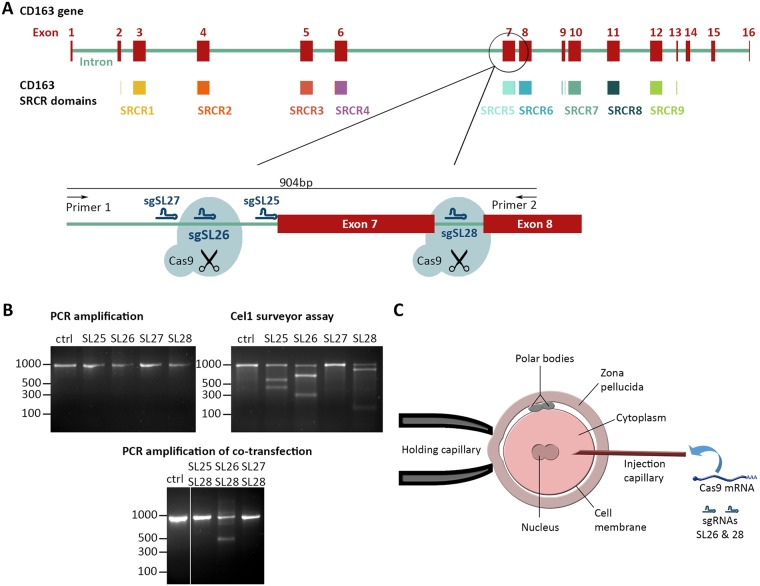
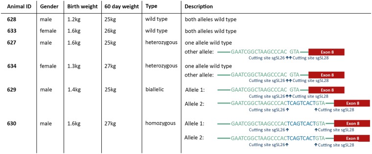
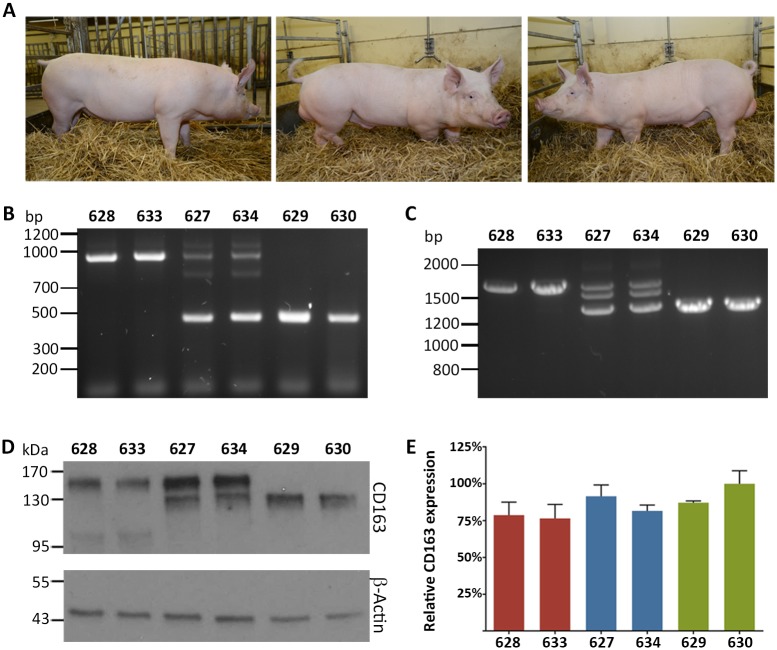
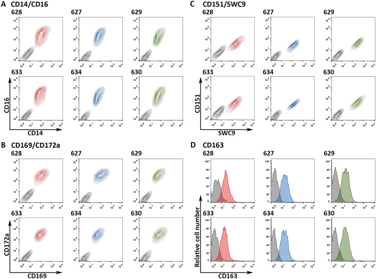
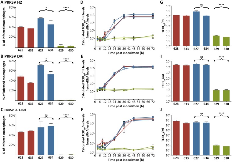
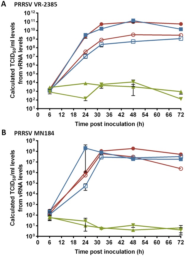
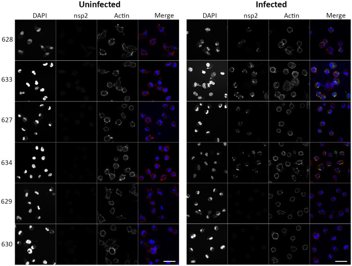
Porcine Reproductive and Respiratory Syndrome (PRRS) is a panzootic infectious disease of pigs, causing major economic losses to the world-wide pig industry. PRRS manifests differently in pigs of all ages but primarily causes late-term abortions and stillbirths in sows and respiratory disease in piglets. The causative agent of the disease is the positive-strand RNA PRRS virus (PRRSV). PRRSV has a narrow host cell tropism, limited to cells of the monocyte/macrophage lineage. CD163 has been described as a fusion receptor for PRRSV, whereby the scavenger receptor cysteine-rich domain 5 (SRCR5) region was shown to be an interaction site for the virus in vitro. CD163 is expressed at high levels on the surface of macrophages, particularly in the respiratory system. Here we describe the application of CRISPR/Cas9 to pig zygotes, resulting in the generation of pigs with a deletion of Exon 7 of the CD163 gene, encoding SRCR5. Deletion of SRCR5 showed no adverse effects in pigs maintained under standard husbandry conditions with normal growth rates and complete blood counts observed. Pulmonary alveolar macrophages (PAMs) and peripheral blood monocytes (PBMCs) were isolated from the animals and assessed in vitro. Both PAMs and macrophages obtained from PBMCs by CSF1 stimulation (PMMs) show the characteristic differentiation and cell surface marker expression of macrophages of the respective origin. Expression and correct folding of the SRCR5 deletion CD163 on the surface of macrophages and biological activity of the protein as hemoglobin-haptoglobin scavenger was confirmed. Challenge of both PAMs and PMMs with PRRSV genotype 1, subtypes 1, 2, and 3 and PMMs with PRRSV genotype 2 showed complete resistance to viral infections assessed by replication. Confocal microscopy revealed the absence of replication structures in the SRCR5 CD163 deletion macrophages, indicating an inhibition of infection prior to gene expression, i.e. at entry/fusion or unpacking stages.


Conflict of interest statement

I have read the journal's policy and the authors of this manuscript have the following competing interests: CB, SGL, TAA, CBAW, and ALA have patent claims on the targeted deletion described in the manuscript. AJM is employed by a commercial company, Genus plc, a commercial livestock breeder.

Figures

Fig 1
Fig 1. Generation of an exon 7 deletion in CD163 using CRISPR/Cas9.
A) Schematic of the CD163 gene in the pig genome on chromosome 5. Indicated in red are the 16 exons encoding the CD163 mRNA, in varied colors underneath are the 9 scavenger receptor cysteine-rich (SRCR) domains that form the pearl on a string structure of the CD163 protein. Excision of exon 7 using two guide RNAs (sgSL26 & sgSL28) located in the flanking introns should result in SRCR 5 removal from the encoded protein. Indicated are also the locations of sgRNAs SL25 and SL27. B) In vitro assessment of guide RNAs sgSL25, sgSL26, sgSL27, and sgSL28. PK15 cells were transfected with either a single plasmid encoding a guide RNA + Cas9 or co-transfected with combination of two such plasmids. Transfected cells were identified by GFP expression and isolated by FACS. Cutting efficiency of single guide RNA transfection was assessed by a Cel1 surveyor assay. Relative efficiency of exon 7 deletion upon double transfection was assessed by PCR. C) Schematic of the Cas9/guide RNA injection into zygotes. The injection mix was injected into the cytoplasm of zygotes and contained uncapped, non-polyadenylated guide RNAs sgSL26 and sgSL28, as well as capped, polyadenylated Cas9 mRNA.
Fig 2
Fig 2. Genotypes and growth of assessed F1 animals.
Fig 3
Fig 3. Excision of exon 7 results in an SRCR5 CD163 deletion in pigs.
A) Representative photos of the male sibling pigs with three different ΔSRCR5 genotypes at 5 months of age. Left, wild type pig 628, middle, heterozygous pig 627, and right, biallelic pig 629. B) Genotyping of pulmonary alveolar macrophages (PAMs). DNA was extracted from PAMs and genotype assessed by PCR across Intron 6 to Exon 8. The unmodified genome PCR is predicted to result in a 900 bp product, whilst exon 7 deletion should result in a 450 bp PCR product. C) RNA phenotype of pulmonary alveolar macrophages. RNA was extracted from PAMs, converted into cDNA using oligo(dT) primer, and analyzed by PCR across Exons 4–9. The unmodified cDNA should result in a 1686 bp product, whilst the exon 7 deletion is expected to yield a 1371 bp product. D) Protein phenotype of CD163 from PAMs. PAM cells were lysed with reducing SDS sample buffer and CD163 expression analyzed by western blot. E) CD163 mRNA levels in PAMs. RNA was extracted from the same number of PAM cells, DNA removed by DNase treatment, and RNA quantified by 1-step RT-qPCR. Expression levels were normalized using β-Actin expression levels and to the highest CD163-expressing animal. Error bars represent SEM, n = 3*2.
Fig 4
Fig 4. ΔSRCR5 peripheral blood monocyte-derived macrophages (PMMs) are fully differentiated and express macrophage-specific markers.
Peripheral blood monocytes were isolated from the blood of the wild type (red), heterozygous (blue), and ΔSRCR5 (green) animals. Following cultivation in the presence of recombinant human CSF1 (rhCSF1) for seven days PMMs were analyzed by FACS. A) Co-staining with CD14-FITC and CD16-PE antibodies recognizing the native structure of the proteins (colored contour plots; red wild type, blue heterozygous, green ΔSRCR5) relative to isotype controls (grey). B) Co-staining with CD169-FITC and CD172a-PE antibodies recognizing the native structure of the proteins (colored contour plots) relative to isotype controls (grey). C) Co-staining with SWC9 (CD203a)-FITC and CD151-RPE antibodies recognizing the native structure of the proteins (colored contour plots) relative to isotype controls (grey). D) Staining against the native structure of surface expressed CD163 (colored) relative to an isotype control staining (grey).
Fig 5
Fig 5. ΔSRCR5 pulmonary alveolar macrophages (PAMs) are not susceptible to infection with PRRSV genotype 1.
A-C) PAMs from wild type (red), heterozygous (blue), and ΔSRCR5 (green) animals were inoculated at MOI = 1 of PRRSV genotype 1, subtype 1 (strain H2, A), subtype 2 (strain DAI, B), and subtype 3 (strain SU1-Bel, C). 19 hpi cells were detached, fixed and stained with an anti PRRSV-N protein antibody and CD163. Infection was quantified by FACS analysis. Over 98% of infected macrophages were qualified as CD163 positive. Infection levels were statistically analyzed using an unpaired t-test of all wild type against all heterozygous or all biallelic / homozygous data points. Error bars represent SEM, n = 3. D-F) Replication growth curves of PRRSV genotype 1, subtype 1 (strain H2, C), subtype 2 (strain DAI, D), and subtype 3 (strain SU1-Bel, F). PAMs from wild type (red, 628 filled circle, 633 open circle), heterozygous (blue, 627 filled square, 633 open square), and ΔSRCR5 (green, 629 triangle pointing down, 630 triangle pointing up) animals were inoculated at MOI = 0.1 of the respective strain. Cell supernatant was collected at indicated time points to measure the released viral RNA by RT-qPCR. Error bars represent SEM, n = 3*2. G-J) Quantification of infectious particles produced at 48 hpi by TCID50 analysis. Cell supernatant collected at the 48 hpi time point of infection of the time-course experiment was analyzed for infectious viral particle production quantified by TCID50. Infection levels were statistically analyzed using an unpaired t-test of all wt against all het or all ΔSRCR5. Error bars represent SEM, n = 3.
Fig 6
Fig 6. ΔSRCR5 peripheral blood monocyte-derived macrophages (PMMs) are not susceptible to infection with PRRSV genotype 2.
Replication of PRRSV on PMMs in long-term infections with genotype 2, A) highly virulent strain VR-2385, and B) highly pathogenic strain MN-184. PMMs from wild type (red, 628 filled circle, 633 open circle), heterozygous (blue, 627 filled square, 633 open square), and ΔSRCR5 (green, 629 triangle pointing down, 630 triangle pointing up) animals were inoculated at MOI = 0.1 of the respective strain. Cell supernatant was collected at indicated time points to measure the released viral RNA by RT-qPCR. Error bars represent SEM, n = 3*2.
Fig 7
Fig 7. PRRSV infection of ΔSRCR5 pulmonary alveolar macrophages (PAMs) is halted prior to the formation of the replication/transcription complex.
PAMs from wild type (top two panels), heterozygous (middle two panels), and ΔSRCR5 (bottom two panels) animals were inoculated at MOI = 2 with PRRSV genotype 1, subtype 3 (strain SU1-Bel). 22 hpi cells were fixed and stained with an anti PRRSV-nsp2 antibody, DAPI, and phalloidin. Scale bar represents 40 μm.
Fig 8
Fig 8. ΔSRCR peripheral blood monocyte-derived macrophages (PMMs) still function as hemoglobin-haptoglobin (Hb-Hp) scavengers.
A) Induction of HO-1 expression by Hb-Hp uptake. PMMs were incubated for 24 h in presence of 100 μg/ml Hb-Hp. RNA was isolated from cells and levels of heme oxygenase 1 (HO-1) mRNA determined by RT-qPCR (outlined bars uninduced, filled bars Hb-Hp uptake induced; red wild type, blue heterozygous, green ΔSRCR5). Expression levels were normalized using β-Actin expression levels and to the level of unstimulated HO-1 mRNA expression of each animal. Uninduced versus induced levels of HO-1 expression were analyzed by an unpaired t-test. Error bars represent SEM, n = 3. B) PMMs were incubated for 24 h in presence of 100 μg/mol Hb-Hp. PMMs were lysed with reducing SDS sample buffer and HO-1 protein expression analyzed by western blot. C) Quantification of Hb-Hp uptake. PMMs were incubated in presence of 10 μg/ml HbAF488-Hp for 30 min. Uptake of HbAF488-Hp was measured by FACS analysis (colored contour plots; red wild type, blue heterozygous, green ΔSRCR5) relative to isotype controls (grey). D) Visualization of Hb-Hp uptake. PMMs were incubated for 30 min with 10 μg/ml HbAF488-Hp. Cells were fixed, permeabilized and stained against CD163 and with DAPI. Representatively shown are pig 628 as wild type and 630 as ΔSRCR5 animals. Scale bar represents 40 μm.

References

    1. Russell P, Atkinson K, Krishnan L (1980) Recurrent reproductive failure due to severe placental villitis of unknown etiology. J Reprod Med 24: 93–98. - PubMed
    1. Wensvoort G, Terpstra C, Pol JM, ter Laak EA, Bloemraad M, et al. (1991) Mystery swine disease in The Netherlands: the isolation of Lelystad virus. Vet Q 13: 121–130. 10.1080/01652176.1991.9694296 - DOI - PubMed
    1. Karniychuk UU, Saha D, Geldhof M, Vanhee M, Cornillie P, et al. (2011) Porcine reproductive and respiratory syndrome virus (PRRSV) causes apoptosis during its replication in fetal implantation sites. Microb Pathog 51: 194–202. 10.1016/j.micpath.2011.04.001 - DOI - PubMed
    1. Pejsak Z, Stadejek T, Markowska-Daniel I (1997) Clinical signs and economic losses caused by porcine reproductive and respiratory syndrome virus in a large breeding farm. Vet Microbiol 55: 317–322. - PubMed
    1. Christianson WT, Joo HS (1994) Porcine reproductive and respiratory syndrome: A review. J Swine Health Prod 2: 10–28.

Publication types

MeSH terms