. 2017 Feb 23;12(2):e0172721.
doi: 10.1371/journal.pone.0172721. eCollection 2017.

Identification of the ER-resident E3 ubiquitin ligase RNF145 as a novel LXR-regulated gene

Affiliations

Identification of the ER-resident E3 ubiquitin ligase RNF145 as a novel LXR-regulated gene

Emma C L Cook et al. PLoS One. .

Abstract

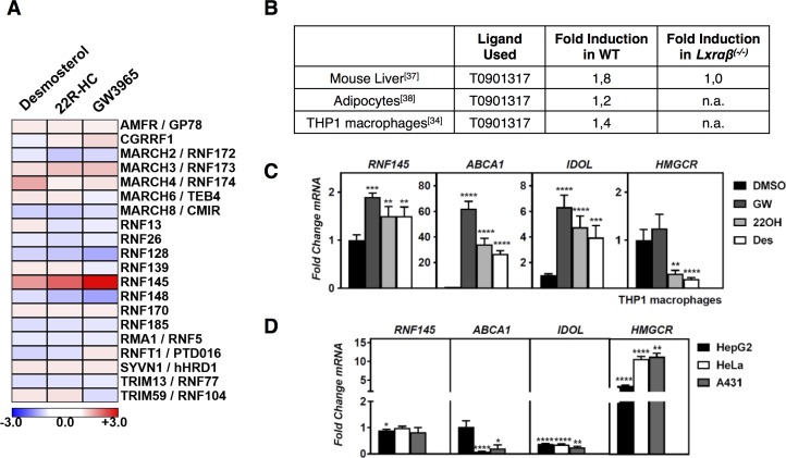
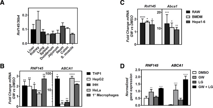
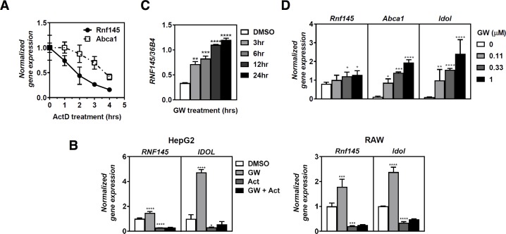
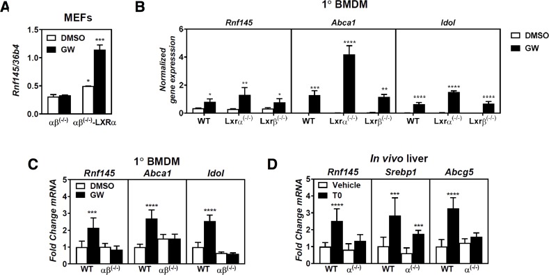
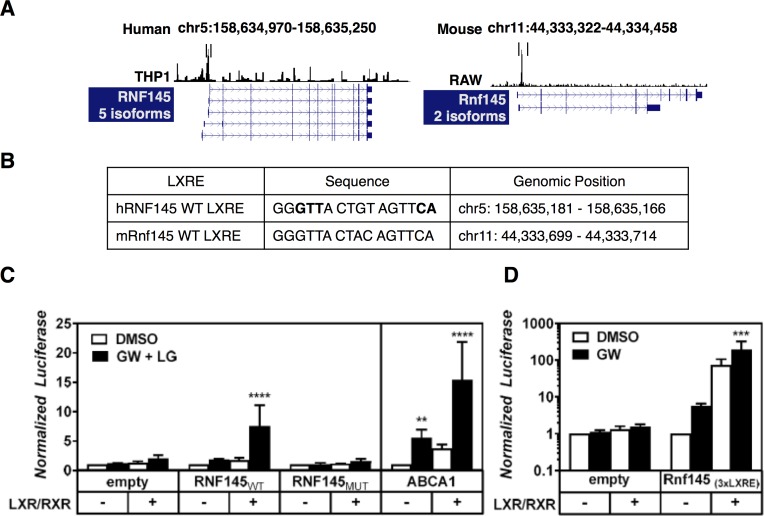
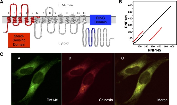
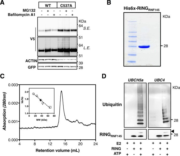
Cellular cholesterol metabolism is subject to tight regulation to maintain adequate levels of this central lipid molecule. Herein, the sterol-responsive Liver X Receptors (LXRs) play an important role owing to their ability to reduce cellular cholesterol load. In this context, identifying the full set of LXR-regulated genes will contribute to our understanding of their role in cholesterol metabolism. Using global transcriptional analysis we report here the identification of RNF145 as an LXR-regulated target gene. We demonstrate that RNF145 is regulated by LXRs in both human and mouse primary cells and cell lines, and in vivo in mice. Regulation of RNF145 by LXR depends on a functional LXR-element in its proximal promotor. Consistent with LXR-dependent regulation of Rnf145 we show that regulation is lost in macrophages and fibroblasts from Lxrαβ(-/-) mice, and also in vivo in livers of Lxrα(-/-) mice treated with the LXR synthetic ligand T0901317. RNF145 is closely related to RNF139/TRC8, an E3 ligase implicated in control of SREBP processing. However, silencing of RNF145 in HepG2 or HeLa cells does not impair SREBP1/2 processing and sterol-responsive gene expression in these cells. Similar to TRC8, we demonstrate that RNF145 is localized to the ER and that it possesses intrinsic E3 ubiquitin ligase activity. In summary, we report the identification of RNF145 as an ER-resident E3 ubiquitin ligase that is transcriptionally controlled by LXR.


Conflict of interest statement

Competing Interests: The authors have declared that no competing interests exist.

Figures

Fig 1
Fig 1. Identification of RNF145 as an LXR target.
(A) Heat map presentation of the expression of predicted ER-resident E3 ubiquitin ligases in THP1 in response to treatment with desmosterol, 22R-HC, and GW3965 [39] (B) CHIP-seq experiments in which an LXR-ligand-responsive peak in the proximal promoter region of RNF145 was detected are shown. The fold-induction by the LXR synthetic ligand, T0901317, that was subsequently determined by transcriptomic analysis is indicated. The response in livers of Lxrα(-/-) was also evaluated; n.a, not available. (C) THP1 cells were grown in sterol-depletion medium and exposed to different LXR ligands for 6 hours: 1μM GW3965 (GW), 5μM 22(R)-hydroxycholesterol (22OH), 5μM Desmosterol (Des), or to vehicle (DMSO). Expression of the indicated genes was determined by qPCR and expressed as fold changes relative to vehicle control. Bars show mean ± SD and significant differences with vehicle are indicated (n = 4). (D) The indicated cells lines were incubated for 16 hours with sterol-depletion medium. Expression of the indicated genes was determined by qPCR and graph expresses fold change of sterol-depleted cells over complete medium. Bars show mean ± SD and significant differences from a value of 1 corresponding to no change (n≥4).
Fig 2
Fig 2. RNF145 is expressed in mouse tissues and is broadly regulated by LXR activation.
(A) Expression of Rnf145 was evaluated in the indicated mouse tissues by qPCR. Bars indicate mean ± SD (n = 3) (B,C) The indicated (B) human and (C) mouse cell lines and primary macrophages were cultured in lipoprotein-depletion medium for 16 hours and subsequently treated for 6 hours with 1μM GW3965 (GW). (D) THP1 cells were cultured in sterol depletion medium for 16 hrs and then treated with 1μM GW3965 (GW) and 100nM LG100268 (LG) for 6 hrs as indicated. Subsequently, ABCA1 and RNF145 expression was determined by qPCR and each bar and error represents the mean fold-change of ligand-treated cells over sterol-depleted cells ± SD (n≥3).
Fig 3
Fig 3. Characterization of ligand-induced expression of RNF145.
(A) RAW264.7 macrophages were treated with 5μg/mL Actinomycin D (ActD) for the indicated time and expression of Abca1 and Rnf145 was determined by qPCR and plotted as mean ± SD relative to untreated cells (n = 3), (B) HepG2 and RAW264.7 cells were treated with 1μM GW3965 for 6 hours in the presence or absence of 5μg/ml actinomycinD for 4 hours, after which expression of the indicated genes was measured by qPCR. Bars indicate mean ± SD (n = 3) (C,D) THP1 macrophages were cultured in sterol-depletion medium for 16 hrs and then treated with (C) 1μM GW3965 (GW) for the indicated time, or (D) with the indicated concentration of GW3965 for 4 hrs. Subsequently, gene expression was evaluated qPCR and bars indicate mean ± SD (n = 3)
Fig 4
Fig 4. LXR-dependent regulation of RNF145.
(A) Mouse embryonic fibroblasts from Lxrαβ(-/-) mice or the same cells with stable overexpression of LXRα, or (B) bone-marrow-derived macrophage from wildtype, Lxrα(-/-), and Lxrβ(-/-) mice were cultured (A,B) in sterol-depletion medium for 16 hrs and subsequently treated with 1μM GW3965 (GW) for 6 hours. Expression of indicated genes was analyzed by qPCR and each bar shows the mean ± SD. Statistical significant differences from (A) Lxrαβ(-/-) cells or (B) control treated cells are shown (n = 3). (C) Bone marrow-derived macrophages were isolated from either wildtype (WT) or Lxrαβ(-/-) mice, cultured in sterol-depletion medium overnight and treated with 1μM GW3965 (GW) for 6 hours. Bars show the expression of the indicated genes relative to vehicle control ± SD (n = 4). (D) Expression of the indicated genes was analysed by qPCR in liver samples from WT and Lxrα(-/-) mice that had either been treated with vehicle control or with 0,015% T0901317 in the diet for 14 days. Bars show mean gene expression relative to vehicle control ± SD (n = 6).
Fig 5
Fig 5. LXRE-dependent regulation of RNF145 expression by LXR.
(A) LXR ChIP-seq experiments in human THP1 cells (GSE28319) and RAW macrophage-like cells (GSE50944) were analyzed and used to identify active LXREs within the Rnf145/Rnf145 loci, as graphically illustrated. (B,C) Genomic location of the identified LXREs. In bold, nucleotides that were mutated to disrupt LXR binding (C) A 1kb genomic region upstream of the transcriptional start site of hRNF145 was cloned into a pGL3basic. The putative LXRE was also mutated as indicated above. The empty, RNF145WT, RNF145MUT, and ABCA1 reporter plasmids were co-transfected with or without RXRα and LXRα expression plasmids in HEK 293T cells. 24 hours post-transfection the cells were treated with 1μM GW3965 (LXR) and 100nM LG100268 (RXR) for 24 hours and measured for luciferase signal (n≥3). (D) Cells were transfected with an empty or a tandem LXRE-containing pGL2 as in C. In all luciferase experiments the transfection efficiency was normalized to co-transfected Renilla luciferase. Bars report normalized chemiluminescence relative to untreated control ± SD (n = 3).
Fig 6
Fig 6. RNF145 is localized to the ER.
(A) The secondary structure of RNF145 (NP_001186312) as predicted by CCTOP is shown. The first five N-terminal transmembrane helices are predicted to contain a sterol-sensing domain [45]. The C-terminal sequence contains a predicted RING structure. (B) An alignment dot plot between RNF139 and RNF145 was generated using PLALIGN [55]. The black line shows similarities between protein sequences. The red lines show repeating sequence motifs within the proteins. (C) HeLa cells were transfected with mRnf145-eGFP (A’,left) and Calnexin mCherry (B’,middle). Merged image is shown (C’, right). Images are representative of three independent experiments.
Fig 7
Fig 7. RNF145 has E3 ubiquitin ligase activity.
(A) HEK293T cells were transfected with wildtype (WT) or RING-mutated (C537A) mRnf145-V5. Subsequently cells were treated with vehicle, 25μM MG-132, or 100nM Bafilomycin A1 for 4 hours. Total cell lysates were immunoblotted as indicated; S.E, short exposure, L.E, long exposure (n = 3) (B) Recombinant His6-RNF145 RING protein was purified and 1 μg loaded on SDS-PAGE gel. An image of a Coomassie Brilliant Blue-stained gel is shown. (C) Size Exclusion Chromatography elution profile of recombinant His6-RNF145 RING protein separated over a Superdex 75 10/300 column calibrated with a set of standard proteins. The elution volume corresponds to an apparent molecular mass of 39 kDa, indicating that in solution His6-RNF145 RING forms a dimer. (D) In vitro ubiquitination assays were done with the RNF145 RING protein in combination with the E2 enzymes UBCH5a and Ubc4. Reactions were carried at 37°C for 2 hours. Subsequently, reactions were immunoblotted as indicated. Arrow indicates an auto-ubiquitylation band of the RING of RNF145. Blots are representative of 2 independent experiments.

Similar articles

Cited by

References

    1. Goldstein JL, DeBose-Boyd RA, Brown MS. Protein sensors for membrane sterols. Cell. 2006;124: 35–46. 10.1016/j.cell.2005.12.022 - DOI - PubMed
    1. Maxfield FR, van Meer G. Cholesterol, the central lipid of mammalian cells. Curr Opin Cell Biol. 2010;22: 422–429. 10.1016/j.ceb.2010.05.004 - DOI - PMC - PubMed
    1. Horton JD, Goldstein JL, Brown MS. SREBPs: activators of the complete program of cholesterol and fatty acid synthesis in the liver. J Clin Invest. 2002;109: 1125–1131. 10.1172/JCI15593 - DOI - PMC - PubMed
    1. Zelcer N, Tontonoz P. Liver X receptors as integrators of metabolic and inflammatory signaling. J Clin Invest. 2006;116: 607–614. 10.1172/JCI27883 - DOI - PMC - PubMed
    1. Radhakrishnan A, Goldstein JL, McDonald JG, Brown MS. Switch-like control of SREBP-2 transport triggered by small changes in ER cholesterol: a delicate balance. Cell Metab. 2008;8: 512–521. 10.1016/j.cmet.2008.10.008 - DOI - PMC - PubMed

MeSH terms