Review
. 2017 Feb 22;93(4):747-765.
doi: 10.1016/j.neuron.2017.01.014.

Neural Circuitry of Wakefulness and Sleep

Affiliations
Review

Neural Circuitry of Wakefulness and Sleep

Thomas E Scammell et al. Neuron. .

Abstract

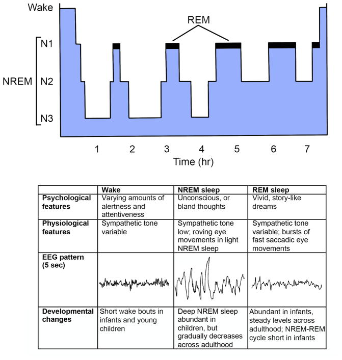
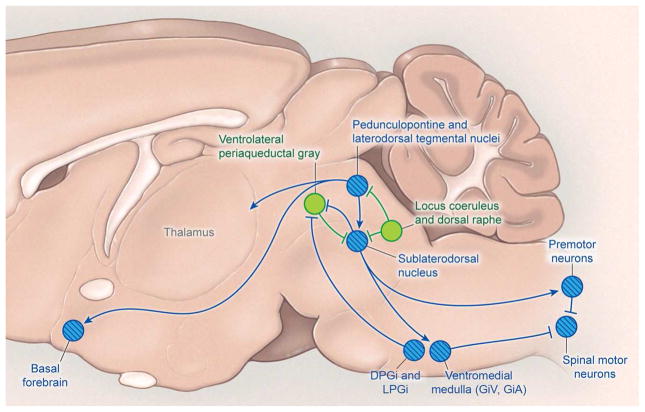
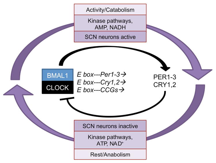
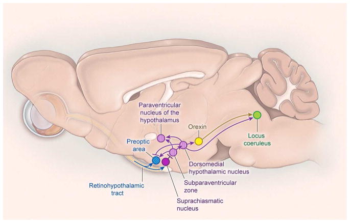
Sleep remains one of the most mysterious yet ubiquitous animal behaviors. We review current perspectives on the neural systems that regulate sleep/wake states in mammals and the circadian mechanisms that control their timing. We also outline key models for the regulation of rapid eye movement (REM) sleep and non-REM sleep, how mutual inhibition between specific pathways gives rise to these distinct states, and how dysfunction in these circuits can give rise to sleep disorders.

Keywords: REM sleep; arousal; chemogenetics; circadian; hypocretin; monoamines; non-REM sleep; optogenetics; orexin; pharmacogenetics; wake.


Conflict of interest statement

Conflicts of Interest: none

Figures

Figure 1
Figure 1. Sleep physiology
A) Over the night, a typical young adult rapidly enters deep NREM sleep (N3) and then cycles between NREM and REM sleep about every 90 minutes. As homeostatic sleep pressure dissipates across the night, NREM sleep become lighter and REM sleep episodes become longer. B) Features of wake, NREM sleep, and REM sleep.
Figure 2
Figure 2. Wake-promoting pathways
A) Several neurochemical systems promote arousal and the fast cortical activity typical of wakefulness. Monoaminergic neurons (light green) in the rostral brainstem and caudal hypothalamus directly innervate the cortex as well as many subcortical regions including the hypothalamus and thalamus. These monoaminergic regions include noradrenergic neurons of the locus coeruleus, serotonergic neurons of the dorsal and median raphe nuclei, dopaminergic neurons of the ventral tegmental area, and histaminergic neurons of the tuberomammillary nucleus. Wake-promoting signals also arise from the parabrachial nucleus and cholinergic regions (dark green with hatching), including the pedunculopontine and laterodorsal tegmental nuclei and basal forebrain.
Figure 3
Figure 3. Projections of the orexin neurons
The orexin neuropeptides are produced by neurons in the lateral hypothalamus and excite neurons in the cortex, midline thalamus, and all wake-promoting brain regions.
Figure 4
Figure 4. NREM sleep-promoting pathways
GABAergic neurons in the ventrolateral preoptic area and median preoptic nucleus promote sleep by inhibiting wake-promoting neurons in the caudal hypothalamus and brainstem. The basal forebrain also contains sleep-active neurons that may promote sleep via projections within the BF and direct projections to the cortex. GABAergic neurons of the parafacial zone (PZ) may promote sleep by inhibiting the parabrachial nucleus. The cortex contains scattered NREM sleep-active neurons that contain both GABA and neuronal nitric oxide synthase (nNOS). Blue circles with hatching denote NREM sleep-promoting nuclei.
Figure 5
Figure 5. REM sleep-promoting pathways
The sublaterodorsal nucleus (SLD) plays a crucial role in regulating REM sleep. Glutamatergic neurons of the SLD produce the muscle paralysis of REM sleep by exciting GABAergic/glycinergic neurons in the ventromedial medulla and spinal cord that hyperpolarize motor neurons. Cholinergic neurons of the pedunculopontine and laterodorsal tegmental nuclei also promote REM sleep and may help drive the fast EEG activity typical of REM sleep. During wake and NREM sleep, the SLD is inhibited by GABAergic neurons of the ventrolateral periaqueductal grey and adjacent lateral pontine tegmentum as well as monoaminergic neurons of the locus coeruleus and raphe nuclei. During REM sleep, the ventrolateral periaqueductal grey is likely inhibited by GABAergic neurons of the SLD and medulla. REM sleep-promoting nuclei are shown in blue with hatching; REM sleep-suppressing nuclei are shown in green. DPGi, dorsal paragigantocellular reticular nucleus; LPGi, lateral paragigantocellular nucleus; GiV, ventral gigantocellular reticular nucleus; GiA, alpha gigantocellular reticular nucleus.
Figure 6
Figure 6. Circadian timekeeping in cells is orchestrated by transcriptional-translational and post-translational feedback loops
The transcription factors BMAL1 and CLOCK form heterodimers that activate the transcription of E box-containing clock-controlled genes (CCGs) including the Period (Per) and Cryptochrome (Cry) genes. Per and Cry gene products subsequently inhibit the activity of BMAL1/CLOCK. Additionally, circadian oscillations in kinase pathways, energy metabolism (AMP/ATP ratio), and redox state (NAD+/NADH ratio) link global control of cellular physiology and metabolism to the molecular clock.
Figure 7
Figure 7. Neural pathways that regulate the circadian timing of sleep and other rhythms
The suprachiasmatic nucleus is the master circadian pacemaker and sits just above the optic chiasm. Neuronal rhythms generated by the SCN are synchronized with the daily light-dark cycle by direct inputs from the retina via the retinohypothalamic tract (RHT). The RHT also has small projections that may influence the activity of sleep-promoting neurons in the preoptic area. Most output signals of the SCN are relayed through the subparaventricular zone (SPZ) and then to the dorsomedial nucleus of the hypothalamus (DMH). The DMH regulates the timing of wakefulness via excitatory projections to the orexin neurons and locus coeruleus and inhibitory projections to the preoptic area. Signals from the SPZ and DMH also regulate the circadian rhythms of heart rate, blood pressure, body temperature, locomotor activity, and feeding. Circadian signals to the paraventricular nucleus of the hypothalamus regulate the daily melatonin rhythm.

References

    1. Adamantidis A, Salvert D, Goutagny R, Lakaye B, Gervasoni D, Grisar T, Luppi PH, Fort P. Sleep architecture of the melanin-concentrating hormone receptor 1-knockout mice. Eur J Neurosci. 2008;27:1793–1800. - PubMed
    1. Adamantidis AR, Zhang F, Aravanis AM, Deisseroth K, de Lecea L. Neural substrates of awakening probed with optogenetic control of hypocretin neurons. Nature. 2007;450:420–424. - PMC - PubMed
    1. Alam MA, Kumar S, McGinty D, Alam MN, Szymusiak R. Neuronal activity in the preoptic hypothalamus during sleep deprivation and recovery sleep. J Neurophysiol. 2014;111:287–299. - PMC - PubMed
    1. Albus H, Vansteensel MJ, Michel S, Block GD, Meijer JH. A GABAergic mechanism is necessary for coupling dissociable ventral and dorsal regional oscillators within the circadian clock. Curr Biol. 2005;15:886–893. - PubMed
    1. Allada R, Siegel JM. Unearthing the phylogenetic roots of sleep. Curr Biol. 2008;18:R670–R679. - PMC - PubMed