. 2017 Feb 23;17(1):154.
doi: 10.1186/s12885-017-3150-7.

Early evaluation of sunitinib for the treatment of advanced gastroenteropancreatic neuroendocrine neoplasms via CT imaging: RECIST 1.1 or Choi Criteria?

Affiliations

Early evaluation of sunitinib for the treatment of advanced gastroenteropancreatic neuroendocrine neoplasms via CT imaging: RECIST 1.1 or Choi Criteria?

Yanji Luo et al. BMC Cancer. .

Abstract

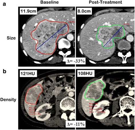
Background: The aim of this study was to assess and compare the Response Evaluation Criteria in Solid Tumors version 1.1 (RECIST 1.1) and the Choi criteria in evaluating the early response of advanced gastroenteropancreatic neuroendocrine neoplasms (GEP-NENs) treated with sunitinib.

Methods: Eighteen patients with pathologically proven advanced GEP-NENs treated with sunitinib were enrolled in the study. Pre- and post-treatment CT scans (plain, biphasic enhanced CT scan) were performed on all patients. Changes in the target tumor size and density from pre-treatment to 1.4-3.1 months after treatment were measured and recorded for each patient. Tumor responses were identified using RECIST 1.1 and Choi criteria. The time to tumor progression (TTP) for each patient was measured and compared between groups using the Kaplan-Meier method.

Results: Among the 18 patients, 4 (22%) exhibited a partial response (PR), 9 (50%) exhibited stable disease (SD), and 5 (28%) experienced progressive disease (PD), using RECIST 1.1. However, based on the Choi criteria, 8 (44%) patients exhibited a PR, 4 (22%) exhibited SD, and 6 (33%) experienced PD. According to RECIST 1.1, the median TTP of PR, SD and PD group were 16.6, 10.8 and 2.3 months, respectively. The TTP of the PR group was significantly longer than that of the PD group (P = 0.007) but insignificant when compared to the SD group (P = 0.131). According to Choi criteria, the median TTP of PR, SD and PD group were not reached, 10.8 and 2.3 months, respectively. The TTP of the PR group was significantly longer than that of the SD (P = 0.026) and PD groups (P < 0.001).

Conclusion: The Choi criteria appear to be more sensitive and more precise than RECIST 1.1 in assessing the early response of advanced GEP-NENs treated with sunitinib.

Keywords: Computed tomography; Gastroenteropancreatic neuroendocrine neoplasms; Sunitinib; Time to tumor progression.


Figures

Fig. 1
Fig. 1
Example of evaluating percentile change in tumor size and density. Pre-treatment and post-treatment CT scans showing shrinkage of hepatic metastases from a pancreatic neuroendocrine tumor in one’s fifties, the percentile change of tumor size was (8.0–11.9)/11.9 × 100% = −33% (a). In another lesion in the same patient, the hepatic metastases appeared more heterogeneous, with high vascularization prior to treatment. Sunitinib induced a large area of tumor hypodensity, suggesting tumor necrosis, the percentile change of tumor attenuation was (108–121)/121 × 100% = −11% (b). In patients with more than one target lesion, the sum of the longest diameters/density of each target lesion in each patient was computed
Fig. 2
Fig. 2
Waterfall plot of the percent change in tumor size (a) and density (b) at the first evaluation after sunitinib treatment
Fig. 3
Fig. 3
A primary pancreatic neuroendocrine tumor with multiple hepatic metastases (G2) in one’s fourties. (a) Pre-treatment CT scan showing a large mass in the pancreatic body with a heterogeneous, hyperdense tumor (white arrow, size: 5.5 cm, density: 91 HU). (b) CT scan obtained 2.8 months after treatment of sunitinib showing that the lesion had become significantly smaller in size and more hypodense (white arrow, size: 2.5 cm, density: 44 HU). The percent change in tumor size and density was 55% and 52%, respectively, which was classified as PR by both the Choi criteria and RECIST. Samples obtained through endoscopic ultrasound-guided fine needle tissue acquisition before treatment showing a large trabecular structure, moderate cell atypia (c, original magnification, ×200, hematoxylin-eosin staining) and intense immunoreactivity for VEGFR2 (d, original magnification, ×200, IHC staining)
Fig. 4
Fig. 4
A pancreatic neuroendocrine tumor (G2) with retroperitoneal lymph node metastases in one’s fifties. The pre-treatment CT scan showed (a) retroperitoneal fusion nodules with a relatively low density (white arrows, size: 2.0 cm, density: 82 HU) in front of the abdominal artery. (b) The nodules exhibited a slight reduction in size and an obvious reduction in density (white arrows, size: 1.9 cm, density: 56 HU) at the first evaluation after treatment with sunitinib. The percent change in tumor size and density was 5.0% and 31.7%, respectively. This patient was classified as PR according to the Choi criteria but as SD based on RECIST
Fig. 5
Fig. 5
Kaplan-Meier analyses of the TTP in the PR, SD and PD groups, as classified according to RECIST 1.1 (a) and the Choi criteria (b)

Similar articles

Cited by

References

    1. Modlin IM, Oberg K, Chung DC, Jensen RT, de Herder WW, Thakker RV, et al. Gastroenteropancreatic neuroendocrine tumours. Lancet Oncol. 2008;9:61–72. doi: 10.1016/S1470-2045(07)70410-2. - DOI - PubMed
    1. Yao JC, Hassan M, Phan A, Dagohoy C, Leary C, Mares JE, et al. One hundred years after “carcinoid”: epidemiology of and prognostic factors for neuroendocrine tumors in 35,825 cases in the United States. J Clin Oncol. 2008;26:3063–72. doi: 10.1200/JCO.2007.15.4377. - DOI - PubMed
    1. Modlin IM, Lye KD, Kidd M. A 5-decade analysis of 13,715 carcinoid tumors. Cancer. 2003;97:934–59. doi: 10.1002/cncr.11105. - DOI - PubMed
    1. Klöppel G. Classification and pathology of gastroenteropancreatic neuroendocrine neoplasms. Endocr Relat Cancer. 2011;18(Suppl 1):S1–16. doi: 10.1530/ERC-11-0013. - DOI - PubMed
    1. de Mestier L, Lardière-Deguelte S, Brixi H, O'Toole D, Ruszniewski P, Cadiot G, et al. Updating the surgical management of peritoneal carcinomatosis in patients with neuroendocrine tumors. Neuroendocrinology. 2015;101:105–11. doi: 10.1159/000371817. - DOI - PubMed

MeSH terms

Supplementary concepts