. 2017 Feb 23;12(1):20.
doi: 10.1186/s13024-017-0160-5.

Tissue-type plasminogen activator exerts EGF-like chemokinetic effects on oligodendrocytes in white matter (re)myelination

Affiliations

Tissue-type plasminogen activator exerts EGF-like chemokinetic effects on oligodendrocytes in white matter (re)myelination

Camille Leonetti et al. Mol Neurodegener. .

Abstract

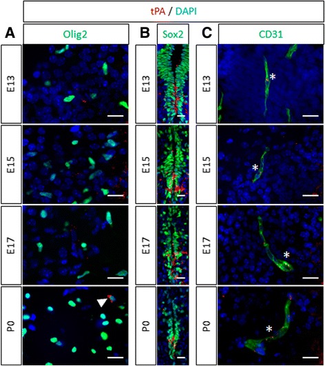
Background: The ability of oligodendrocyte progenitor cells (OPCs) to give raise to myelin forming cells during developmental myelination, normal adult physiology and post-lesion remyelination in white matter depends on factors which govern their proliferation, migration and differentiation. Tissue plasminogen activator (tPA) is a serine protease expressed in the central nervous system (CNS), where it regulates cell fate. In particular, tPA has been reported to protect oligodendrocytes from apoptosis and to facilitate the migration of neurons. Here, we investigated whether tPA can also participate in the migration of OPCs during CNS development and during remyelination after focal white matter lesion.

Methods: OPC migration was estimated by immunohistological analysis in spinal cord and corpus callosum during development in mice embryos (E13 to P0) and after white matter lesion induced by the stereotactic injection of lysolecithin in adult mice (1 to 21 days post injection). Migration was compared in these conditions between wild type and tPA knock-out animals. The action of tPA was further investigated in an in vitro chemokinesis assay.

Results: OPC migration along vessels is delayed in tPA knock-out mice during development and during remyelination. tPA enhances OPC migration via an effect dependent on the activation of epidermal growth factor receptor.

Conclusion: Endogenous tPA facilitates the migration of OPCs during development and during remyelination after white matter lesion by the virtue of its epidermal growth factor-like domain.

Keywords: Development; Endothelial cells; Epidermal growth factor; Lysolecithin; Multiple sclerosis; Myelin; Spinal cord; Vasophilic migration.


Figures

Fig. 1
Fig. 1
Migration of OPC is delayed in tPA −/− mice during spinal cord development. a Photomicrographs from spinal cord sections show representative images (from 3–4 mice) of Olig2 immunostaining (red) and DAPI staining (blue) in the spinal cord (right ventral horn) of WT (left column) and tPA−/− (right column) mice at E13, E15, E17 and P0. b Corresponding quantifications of Olig2+ oligodendrocytes (mean ± SEM, n = 3–4 per group; *p < 0.05). c Photomicrographs from tissue sections show representative images (from 3–4 mice) of Olig2 immunostaining (red) and DAPI staining (blue) in pMN domain of WT (left column) and tPA−/− (right column) mice at E13, E15, E17 and P0. d Corresponding quantifications of Olig2+ oligodendrocytes (mean ± SEM, n = 3–4 per group; *p < 0.05). e Quantification of the percentage of proliferating OLs (Ki67+/Olig2+) in the spinal cord parenchyma (right ventral horn and pMN domain) (mean ± SEM, n = 3–4 per group). E: embryonic day; ec: ependymal canal; ND: not detected; OL: oligodendrocyte; P: postnatal day; pMN domain: motor neuron progenitor domain; RVH: right ventral horn; WT: wild type. tPA −/− : tPA Knock-out. Scale bars: 20 μm
Fig. 2
Fig. 2
Migration of OPC is delayed in tPA −/− mice during corpus callosum development. a Photomicrographs from tissue sections show representative images (from 3 mice) of Olig2 immunostaining (red) and DAPI staining (blue) in the telencephalon of WT (left column) and tPA−/− (right column) mice during corpus callosum development at E15, E17, P0 and P3. Dotted lines show corpus callosum limits. b Corresponding quantifications of Olig2+ oligodendrocytes (mean ± SEM, n = 3 per group; *p < 0.05). E: embryonic day; OL: oligodendrocyte; P: postnatal day; WT: wild type. Scale bars: 100 μm
Fig. 3
Fig. 3
tPA expression during spinal cord development. Photomicrographs from spinal cord sections show representative images (from 3 mice) of tPA (red) with (a) Olig2 (green), (b) Sox2 (green), and (c) CD31 (right column, green) immunostaining in the spinal cord of wild type mice at E13, E15, E17 and P0. Asterisks show colocalization of tPA and CD31 immunoreactivity in endothelial cells. Filled arrowhead shows colocalization of tPA and Olig2 immunoreactivity oligodendrocytes. E: embryonic day; P: postnatal day. Scale bars: 20 μm
Fig. 4
Fig. 4
tPA promotes oligodendrocyte migration along vessels during development at times of intense migration. a Photomicrographs from spinal sections show representative images (from 3 mice) of tPA (red), CD31 (green) and Olig2 (grey) immunostaining in the right ventral horn of WT mice spinal cords at E13. Right image is a 3D reconstruction from confocal acquisitions (on the left). b Photomicrographs from tissue sections show representative images (from 3 mice per group) of Olig2 (red), CD31 (green), and DAPI (blue) immunostaining in the right ventral horn of WT and tPA−/− mice spinal cords at E13. c Quantification of the percentage of Olig2+ oligodendrocytes close (<10 μm) to vessels in the ventral spinal cord parenchyma outside the pMN domain in WT and tPA−/− mice (mean ± SEM, n = 3 per group; *p < 0.05). d Quantification of the percentage of CD31+ staining area in the ventral spinal cord parenchyma outside the pMN domain in WT and tPA−/− mice (mean ± SEM, n = 3 per group; *p < 0.05). E: embryonic day; OL: oligodendrocyte; pMN domain: motor neuron progenitor domain; WT: wild type. Scale bars: 20 μm
Fig. 5
Fig. 5
Remyelination is delayed in tPA −/− after white matter damage in adult mice. a Representative high resolution T2 images and b quantification of the lesion size at 1, 3, 7, 14 and 21 days after lysolecithin injection (dpi) in the corpus callosum of WT and tPA−/− mice. (mean ± SEM, n = 5 per group; *p < 0.05 and **p < 0.01 significantly, Mann–Whitney U test). c Photomicrographs from corpus callosum sections (perilesion) show representative images (from 3 mice) of Olig2 immunostaining (red) and DAPI staining (blue) from WT (left) and tPA−/− (right) mice at 3 days post injection (dpi). (Scale bars: 20 μm). d Corresponding quantifications of Olig2+ oligodendrocytes (mean ± SEM, n = 3 per group; *p < 0.05). e Quantification of the percentage of proliferating OLs (Ki67+Olig2+/Olig2+) in the lesion of WT and tPA−/− mice, 3 days post-injection. (mean ± SEM, n = 3 per group). f Photomicrographs from brain sections (corpus callosum lesion) show representative images (from 3 mice per group) of MBP immunostaining (red) in the ipsilateral and contralateral corpus callosum of WT and tPA−/− mice, 14 and 21 days post-injection (dpi). Dotted lines show quantified area limits. (Scale bars: 100 μm). g Corresponding quantifications of MBP fluorescence intensity (mean ± SEM, n = 3 per group; *p < 0.05). AU: arbitrary unit; dpi: days post injection; OL: oligodendrocyte; WT: wild type. tPA −/− : tPA Knock-out
Fig. 6
Fig. 6
tPA expression after white matter damage in wild type mice. Photomicrographs from tissue sections show representative images of tPA (red), GFAP (green, first column), Iba1 (green, second column), CD31 (green, third column), Olig2 (green, fourth column) and DAPI staining (blue) immunoreactivities in the remyelinating corpus callosum of WT mice, 1, 3, 7, 14 and 21 days after lysolecithin injection. Asterisks show colocalization of tPA and CD31 immunoreactivities in endothelial cells. Filled arrowheads show colocalization of tPA and Olig2 immunoreactivities in oligogendrocytes. (Representative images, n = 3 per group). dpi: days post injection. Scale bars: 20 μm
Fig. 7
Fig. 7
tPA promotes oligodendrocyte migration along vessels after white matter damage. a Photomicrographs from brain sections show representative images (from 3 mice) of tPA (red), CD31 (green) and Olig2 (grey) immunostaining in the corpus callosum lesion of WT mice at 3 days post injection (3 dpi). Right image is a rotation of 3D reconstruction from confocal acquisitions (on the left). b Photomicrographs from tissue sections show representative images (from 3 mice per group) of Olig2 (red), CD31 (green), and DAPI (blue) immunostaining in the corpus callosum (perilesion) of WT and tPA−/− mice spinal cords at 3 days post injection (3 dpi). c Quantification of the percentage of Olig2+ oligodendrocytes close (<10 μm) to vessels in the in the corpus callosum (perilesion) in WT and tPA−/− mice (mean ± SEM, n = 3 per group; *p < 0.05). d Quantification of the percentage of CD31+ staining area in corpus callosum (perilesion) in WT and tPA−/− mice (mean ± SEM, n = 3 per group; *p < 0.05). E: embryonic day; OL: oligodendrocyte; pMN domain: motor neuron progenitor domain; WT: wild type. Scale bars: 10 μm
Fig. 8
Fig. 8
tPA induces chemokinesis on cultured OLs. a Rat primary cultures of OPCs were seeded on the upper compartment of Boyden chambers and treated in indicated conditions for 24 h. Photomicrographs show representative fields of the lower compartment after Olig2 immunostaining (green; representative image, n = 9, three independent cultures). b Oligodendrocyte chemokinesis was estimated by counting cells in the lower compartment in control conditions or in the presence of 0.2 μg/mL FGF2, 0.1-10 μg/mL tPA or tPA vehicle. (mean ± SEM; n = 9, three independent cultures; *p < 0.05 and **p < 0.01). c OL proliferation (percentage) during 24 h was estimated by measuring mitochondrial activity with WST-1 test after treatment or not. Control is the basal OL proliferation during 24 h without treatment (mean + SEM; n = 15, five independent cultures). OPC: oligodendrocyte precursor cell; ns: not significant. Scale bars: 100 μm
Fig. 9
Fig. 9
tPA induces chemokinesis on OLs via its EGF-like domain. a Photomicrographs from WT mice tissue sections show representative images of tPA (red), EGFR (green) and Olig2 (blue) immunoreactivities in the ventral ventricular zone of mice spinal cords at E13, E15, E17, P0, P3 and P7. Dotted lines show ependymal canal limits. (Representative images, n = 3 per group). b Photomicrographs from WT mice tissue sections show representative images of EGFR (green) and Olig2 (blue) immunoreactivities in the pMN domain of mice spinal cords at E13. The inlet shows 3D reconstruction from confocal acquisition. (Representative images, n = 3 per group). c Photomicrographs from WT mice tissue sections show representative images of tPA (red), EGFR (green) and Olig2 (blue) immunoreactivities in the contralateral (left column) and ipsilateral (right column) corpus callosum of adult mice, 3, 7, 14 and 21 days after lysolecithin injection. Full lines show lateral ventricle limits and dotted lines show corpus callosum limits. (Representative images, n = 3 per group). d Photomicrographs from adult WT mice tissue sections show representative images of EGFR (green) and Olig2 (blue) immunoreactivities in the lesion 3 days after injection. (Representative images, n = 3 per group). e Rat OPCs were cultured for 24 h. Photomicrographs show representative fields after Olig2 (red) and EGFR (green) immunostaining or the merged images (yellow). (representative images, n = 3 per group). f Rat primary cultures of OPCs were seeded on the upper compartment of Boyden chambers and treated in indicated conditions for 24 h. g Proteins from cultured OPCs treated with tPA (10μg/ml) or corresponding vehicle for indicated times were subjected to western blot for the phosphorylated (P-Erk) or total (Total Erk) forms of Erk. Chemokinesis was estimated by counting cells in the lower compartment in control conditions or in the presence of 0.2 μg/mL FGF2, 10 μg/mL tPA, 10 μg/mL tPA + 5 μM inhibitor of the kinase activity of EGFR (AG1478), 5 μM AG1478, 10 μg/mL tPA complexed with the proteolytic inhibitory peptide GGACK. (mean ± SEM; n = 9, three independent cultures; *p < 0.05, Mann–Whitney U test). cc: corpus callosum; dpi: days post injection; E: embryonic day; LV: lateral ventricle; OPC: oligodendrocyte precursor cell; P: postnatal day. Scale bars: 20 μm

Similar articles

Cited by

References

    1. Franklin RJM, ffrench-Constant C. Remyelination in the CNS: from biology to therapy. Nat Rev Neurosci. 2008;9:839–55. doi: 10.1038/nrn2480. - DOI - PubMed
    1. Goldschmidt T, Antel J, König FB, Brück W, Kuhlmann T. Remyelination capacity of the MS brain decreases with disease chronicity. Neurology. 2009;72:1914–21. doi: 10.1212/WNL.0b013e3181a8260a. - DOI - PubMed
    1. Patrikios P, Stadelmann C, Kutzelnigg A, Rauschka H, Schmidbauer M, Laursen H, et al. Remyelination is extensive in a subset of multiple sclerosis patients. Brain. 2006;129:3165–72. doi: 10.1093/brain/awl217. - DOI - PubMed
    1. Bramow S, Frischer JM, Lassmann H, Koch-Henriksen N, Lucchinetti CF, Sørensen PS, et al. Demyelination versus remyelination in progressive multiple sclerosis. Brain. 2010;133:2983–98. doi: 10.1093/brain/awq250. - DOI - PubMed
    1. Kremer D, Göttle P, Hartung H-P, Küry P. Pushing forward: remyelination as the New frontier in CNS diseases. Trends Neurosci. 2016;39:246–63. doi: 10.1016/j.tins.2016.02.004. - DOI - PubMed

Publication types

MeSH terms

Substances