

Epitopes and Mechanism of Action of the Clostridium difficile Toxin A-Neutralizing Antibody Actoxumab
- PMID: 28232034
- PMCID: PMC7373199
- DOI: 10.1016/j.jmb.2017.02.010
Epitopes and Mechanism of Action of the Clostridium difficile Toxin A-Neutralizing Antibody Actoxumab
Abstract
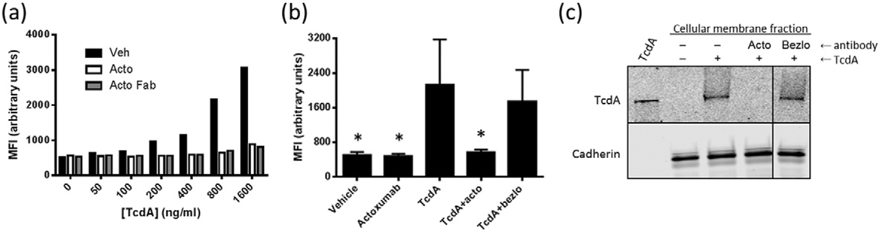
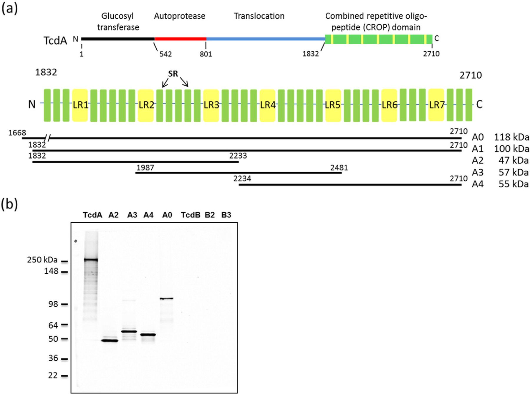
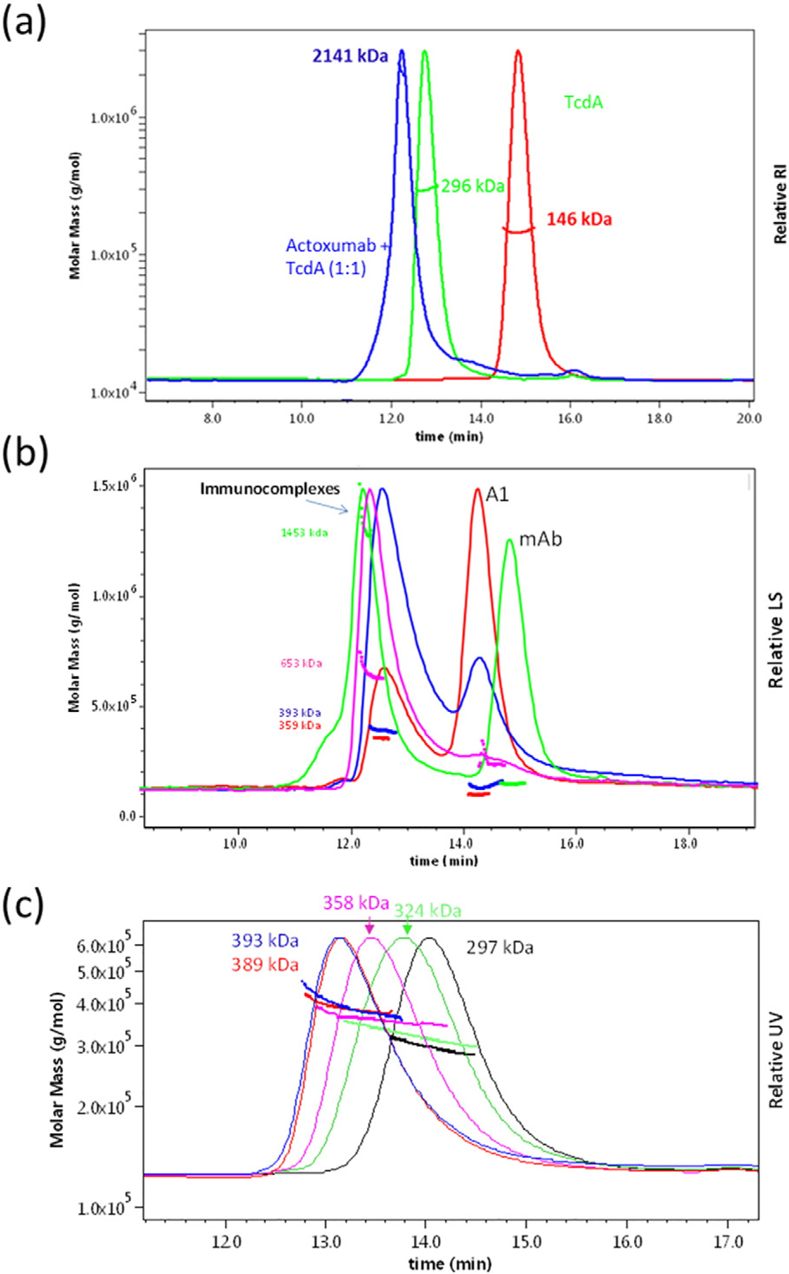
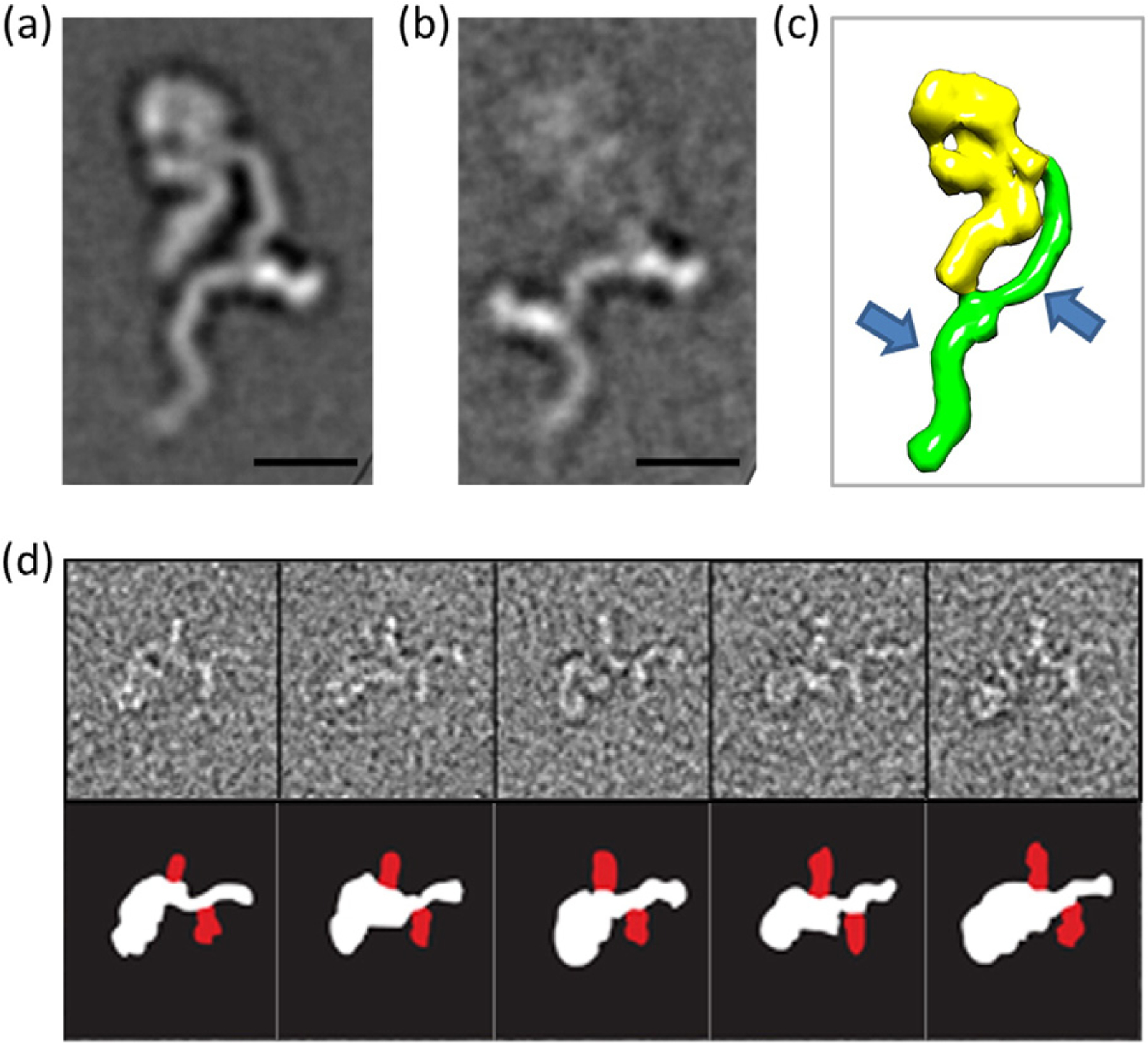
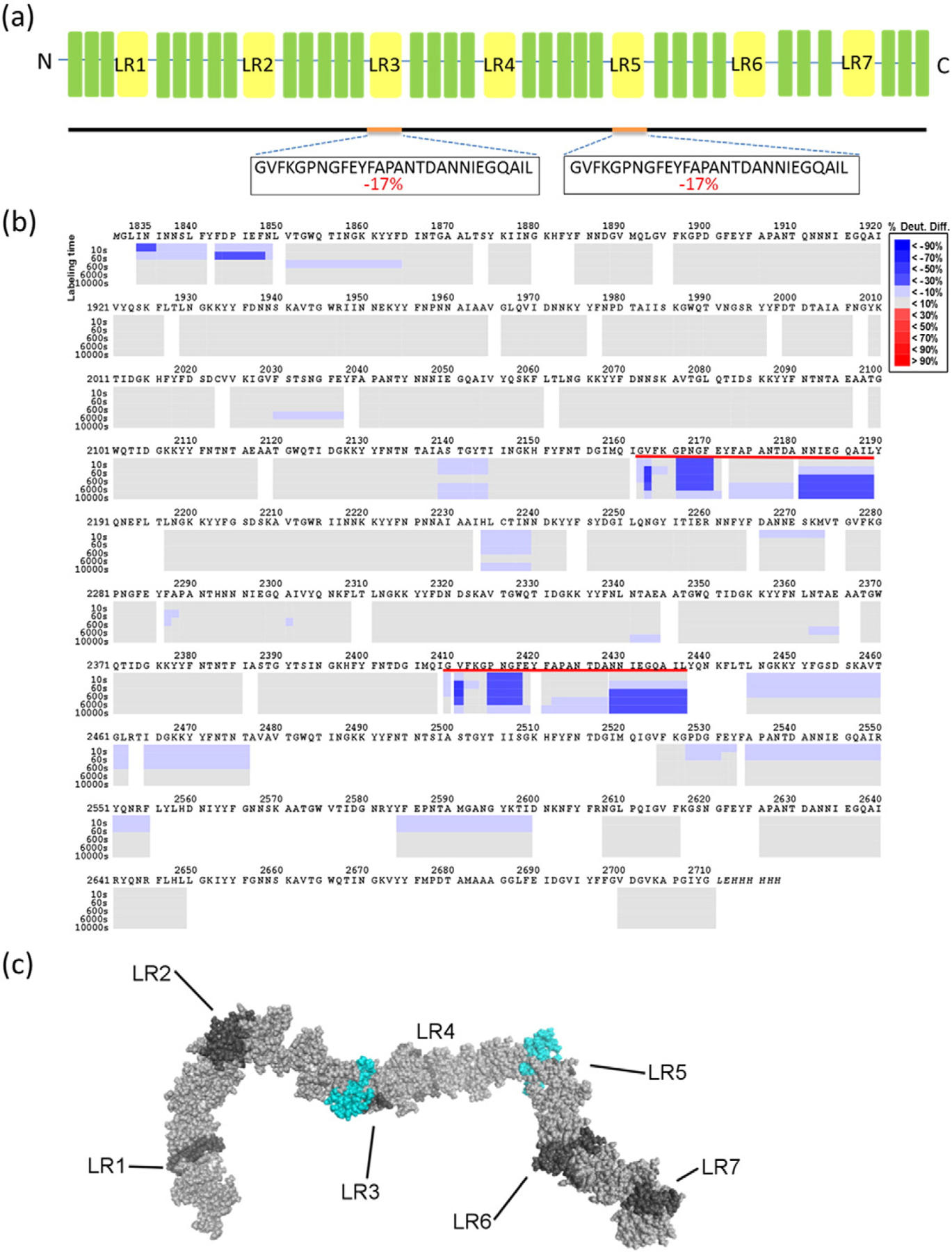
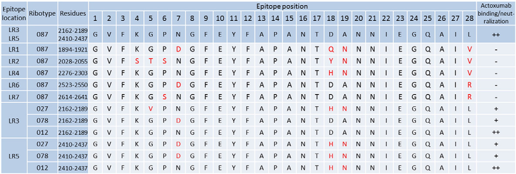
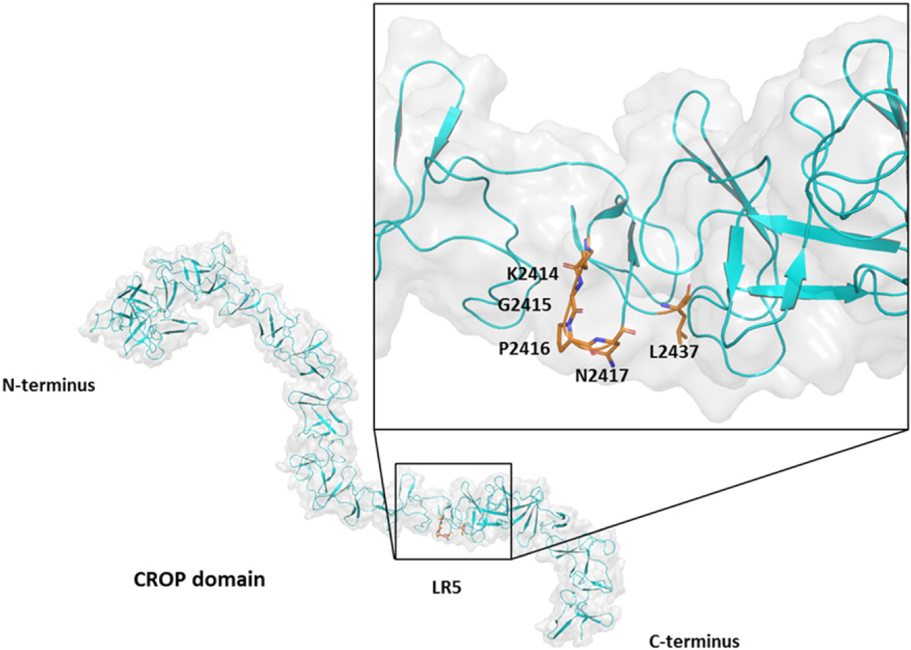
The exotoxins toxin A (TcdA) and toxin B (TcdB) are produced by the bacterial pathogen Clostridium difficile and are responsible for the pathology associated with C. difficile infection (CDI). The antitoxin antibodies actoxumab and bezlotoxumab bind to and neutralize TcdA and TcdB, respectively. Bezlotoxumab was recently approved by the FDA for reducing the recurrence of CDI. We have previously shown that a single molecule of bezlotoxumab binds to two distinct epitopes within the TcdB combined repetitive oligopeptide (CROP) domain, preventing toxin binding to host cells. In this study, we characterize the binding of actoxumab to TcdA and examine its mechanism of toxin neutralization. Using a combination of approaches including a number of biophysical techniques, we show that there are two distinct actoxumab binding sites within the CROP domain of TcdA centered on identical amino acid sequences at residues 2162-2189 and 2410-2437. Actoxumab binding caused the aggregation of TcdA especially at higher antibody:toxin concentration ratios. Actoxumab prevented the association of TcdA with target cells demonstrating that actoxumab neutralizes toxin activity by inhibiting the first step of the intoxication cascade. This mechanism of neutralization is similar to that observed with bezlotoxumab and TcdB. Comparisons of the putative TcdA epitope sequences across several C. difficile ribotypes and homologous repeat sequences within TcdA suggest a structural basis for observed differences in actoxumab binding and/or neutralization potency. These data provide a mechanistic basis for the protective effects of the antibody in vitro and in vivo, including in various preclinical models of CDI.
Keywords: Clostridium difficile infection; TcdA; epitope mapping; monoclonal antibody; toxin neutralization.
Copyright © 2017 Elsevier Ltd. All rights reserved.
Figures
Similar articles
-
Mechanism of action and epitopes of Clostridium difficile toxin B-neutralizing antibody bezlotoxumab revealed by X-ray crystallography.J Biol Chem. 2014 Jun 27;289(26):18008-21. doi: 10.1074/jbc.M114.560748. Epub 2014 May 12. J Biol Chem. 2014. PMID: 24821719 Free PMC article.
-
Broad coverage of genetically diverse strains of Clostridium difficile by actoxumab and bezlotoxumab predicted by in vitro neutralization and epitope modeling.Antimicrob Agents Chemother. 2015 Feb;59(2):1052-60. doi: 10.1128/AAC.04433-14. Epub 2014 Dec 1. Antimicrob Agents Chemother. 2015. PMID: 25451052 Free PMC article. Clinical Trial.
-
Mechanisms of protection against Clostridium difficile infection by the monoclonal antitoxin antibodies actoxumab and bezlotoxumab.Infect Immun. 2015 Feb;83(2):822-31. doi: 10.1128/IAI.02897-14. Epub 2014 Dec 8. Infect Immun. 2015. PMID: 25486992 Free PMC article.
-
Actoxumab + bezlotoxumab combination: what promise for Clostridium difficile treatment?Expert Opin Biol Ther. 2018 Apr;18(4):469-476. doi: 10.1080/14712598.2018.1452908. Epub 2018 Mar 15. Expert Opin Biol Ther. 2018. PMID: 29534621 Review.
-
Bezlotoxumab for the treatment of Clostridium difficile-associated diarrhea.Drugs Today (Barc). 2017 Jul;53(7):385-392. doi: 10.1358/dot.2017.53.7.2655240. Drugs Today (Barc). 2017. PMID: 28837182 Review.
Cited by
-
New treatment approaches for Clostridioides difficile infections: alternatives to antibiotics and fecal microbiota transplantation.Gut Microbes. 2024 Jan-Dec;16(1):2337312. doi: 10.1080/19490976.2024.2337312. Epub 2024 Apr 9. Gut Microbes. 2024. PMID: 38591915 Free PMC article. Review.
-
Neutralizing epitopes on Clostridioides difficile toxin A revealed by the structures of two camelid VHH antibodies.Front Immunol. 2022 Nov 16;13:978858. doi: 10.3389/fimmu.2022.978858. eCollection 2022. Front Immunol. 2022. PMID: 36466927 Free PMC article.
-
Human α-Defensin-6 Neutralizes Clostridioides difficile Toxins TcdA and TcdB by Direct Binding.Int J Mol Sci. 2022 Apr 19;23(9):4509. doi: 10.3390/ijms23094509. Int J Mol Sci. 2022. PMID: 35562899 Free PMC article.
-
Clostridium difficile PCR Cycle Threshold Predicts Free Toxin.J Clin Microbiol. 2017 Sep;55(9):2651-2660. doi: 10.1128/JCM.00563-17. Epub 2017 Jun 14. J Clin Microbiol. 2017. PMID: 28615471 Free PMC article.
-
Functional defects in Clostridium difficile TcdB toxin uptake identify CSPG4 receptor-binding determinants.J Biol Chem. 2017 Oct 20;292(42):17290-17301. doi: 10.1074/jbc.M117.806687. Epub 2017 Aug 23. J Biol Chem. 2017. PMID: 28842504 Free PMC article.
References
-
- Rupnik M, Wilcox MH, Gerding DN, Clostridium difficile infection: new developments in epidemiology and pathogenesis, Nat. Rev. Microbiol 7 (2009) 526–536. - PubMed
-
- Bassetti M, Villa G, Pecori D, Arzese A, Wilcox M, Epidemiology, diagnosis and treatment of Clostridium difficile infection, Expert Rev. Anti-Infect. Ther 10 (2012) 1405–1423. - PubMed
-
- Carter GP, Rood JI, Lyras D, The role of toxin A and toxin B in the virulence of Clostridium difficile, Trends Microbiol. 20 (2012) 21–29. - PubMed
MeSH terms
Substances
Grants and funding
LinkOut - more resources
Full Text Sources
Other Literature Sources
