. 2017 May 1:117:292-304.
doi: 10.1016/j.neuropharm.2017.02.019. Epub 2017 Feb 22.

Increased thrombospondin-4 after nerve injury mediates disruption of intracellular calcium signaling in primary sensory neurons

Affiliations

Increased thrombospondin-4 after nerve injury mediates disruption of intracellular calcium signaling in primary sensory neurons

Yuan Guo et al. Neuropharmacology. .

Abstract

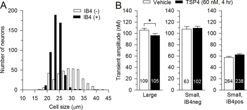
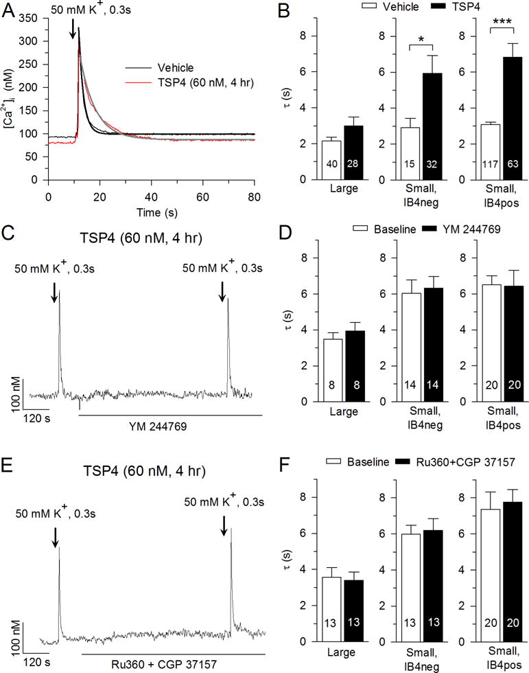
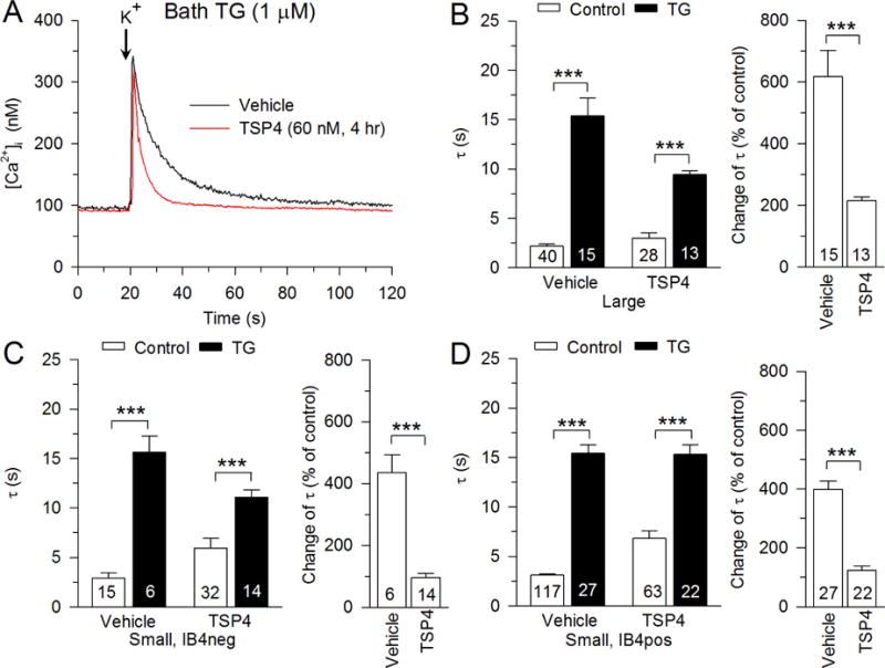
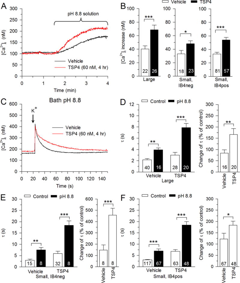
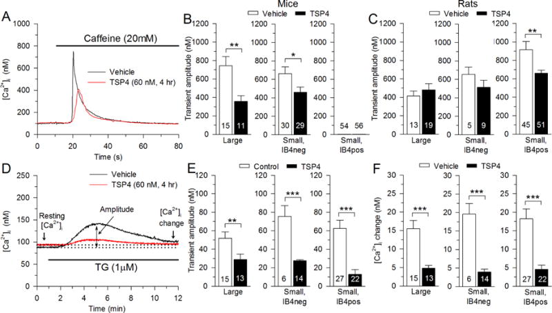
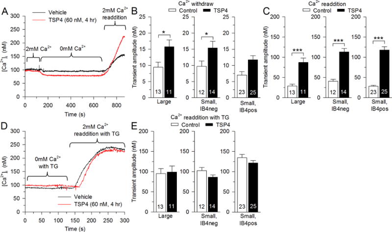
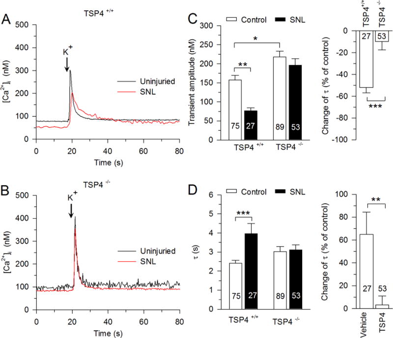
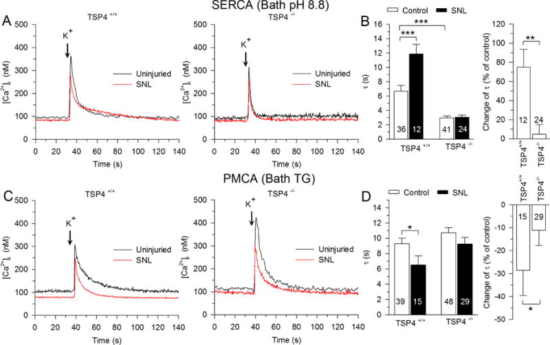
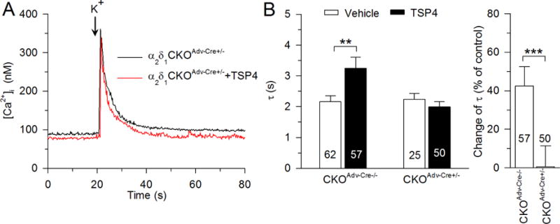
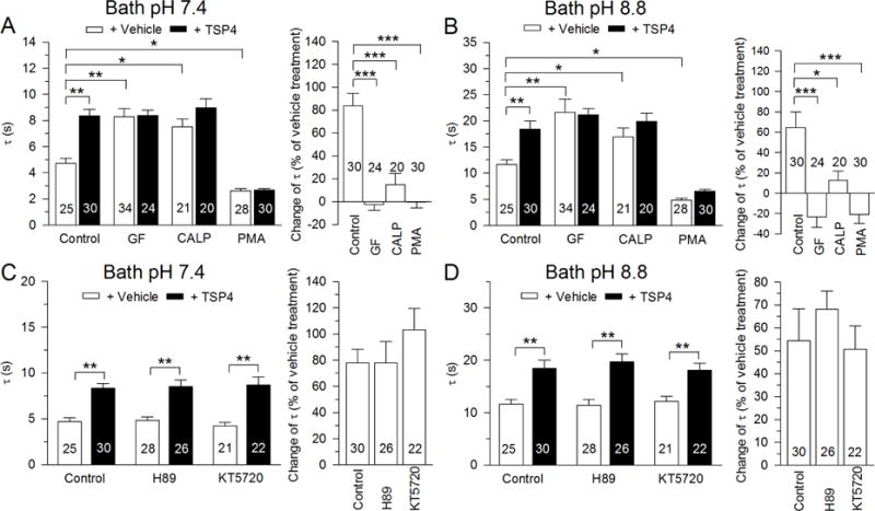
Painful nerve injury disrupts Ca2+ signaling in primary sensory neurons by elevating plasma membrane Ca2+-ATPase (PMCA) function and depressing sarco-endoplasmic reticulum Ca2+-ATPase (SERCA) function, which decreases endoplasmic reticulum (ER) Ca2+ stores and stimulates store-operated Ca2+ entry (SOCE). The extracellular matrix glycoprotein thrombospondin-4 (TSP4), which is increased after painful nerve injury, decreases Ca2+ current (ICa) through high-voltage-activated Ca2+ channels and increases ICa through low-voltage-activated Ca2+ channels in dorsal root ganglion neurons, which are events similar to the effect of nerve injury. We therefore examined whether TSP4 plays a critical role in injury-induced disruption of intracellular Ca2+ signaling. We found that TSP4 increases PMCA activity, inhibits SERCA, depletes ER Ca2+ stores, and enhances store-operated Ca2+ influx. Injury-induced changes of SERCA and PMCA function are attenuated in TSP4 knock-out mice. Effects of TSP4 on intracellular Ca2+ signaling are attenuated in voltage-gated Ca2+ channel α2δ1 subunit (Cavα2δ1) conditional knock-out mice and are also Protein Kinase C (PKC) signaling dependent. These findings suggest that TSP4 elevation may contribute to the pathogenesis of chronic pain following nerve injury by disrupting intracellular Ca2+ signaling via interacting with the Cavα2δ1 and the subsequent PKC signaling pathway. Controlling TSP4 mediated intracellular Ca2+ signaling in peripheral sensory neurons may be a target for analgesic drug development for neuropathic pain.

Keywords: Ca(2+) stores and stimulates store-operated Ca(2+) entry; Intracellular calcium signaling; Neuropathic pain; Plasma membrane Ca(2+)-ATPase; Sarco-endoplasmic reticulum Ca(2+)-ATPase; Thrombospondin-4.


Figures

Fig. 1
Fig. 1
Effects of TSP4 on resting [Ca2+]i of DRG neurons. A, Classification of neurons according to size and IB4 staining. B, Summary data show effects of TSP4 incubation on resting [Ca2+] i in neurons of different groups categorized by size and IB4 binding. The numbers in the bars represent the n for neurons in groups, and bars show mean ± SEM, here and in subsequent figures. * p < 0.05.
Fig. 2
Fig. 2
Effects of TSP4 on the depolarization-induced Ca2+ transient. A, Sample traces show the influence of TSP4 on recovery of the Ca2+ transient induced by neuronal depolarization with KCl (50 mM K+ for 0.3 s). Black and gray lines show single exponential fit of the recovery of [Ca2+] i. B, Summary data show TSP4 decreased [Ca2+] i recovery following depolarization in small-sized neurons. C, Sample trace shows the effect of NCX inhibitor, YM 244769 (5 μM), on recovery of the Ca2+ transient induced by neuronal depolarization with KCl solution. D, Summary data show that YM 244769 did not change [Ca2+] i recovery following depolarization (paired t-test). E, Sample trace shows the effect of mitochondrial Ca2+ uniporter inhibitor, Ru360 (10 μM), and mitochondrial NCX inhibitor, CGP 37157 (10 μM), on recovery of the Ca2+ transient induced by neuronal depolarization with KCl solution. F, Summary data show that Ru360 and CGP 37157 did not change [Ca2+] i recovery following depolarization (paired t-test). * p < 0.05, *** p < 0.001.
Fig. 3
Fig. 3
Regulation of PMCA function by TSP4. A, Sample traces show the effect of TSP4 on PMCA function, revealed by measuring recovery of the depolarization-induced transient during inhibition of SERCA function with thapsigargin (TG, 1 μM). Summary data show that effects of TSP4 on PMCA functions in large-sized neurons (B), small-sized IB4neg neurons (C), and small-sized IB4pos neurons (D). Control data are from Fig. 2. *** p < 0.001.
Fig. 4
Fig. 4
Regulation of SERCA function by TSP4. A, Sample traces show the effect of inhibition of PMCA function with pH 8.8 Tyrode’s solution on neurons with and without TSP4 treatment. B, Summary data show effects of TSP4 on resting [Ca2+] i in pH 8.8 Tyrode’s solution. C, Sample traces show the effect of TSP4 on SERCA function, revealed by measuring recovery of the depolarization-induced transient recovery during inhibition of PMCA function with pH 8.8 Tyrode’s solution. Summary data show that TSP4 decreased SERCA functions in large-sized neurons (D), small-sized IB4neg neurons (E), and small-sized IB4pos neurons (F). Control data are from Fig. 2. * p < 0.05, ** p < 0.01, *** p < 0.001.
Fig. 5
Fig. 5
Regulation of ER Ca2+ store by TSP4. A, Sample traces show the effect of TSP4 on the Ca2+ transient induced by release of ER stores by caffeine (20 mM). B, Summary data show reduced releasable Ca2+ stores, measured as Ca2+ transient amplitude, in mouse neurons incubated in TSP4. C, Summary data show TSP4 effects in rat neurons. D, Sample traces show the effect of TSP4 on the Ca2+ transient induced by the release of stores by the SERCA blocker TG, 1 μM. E, Summary data show TSP4 suppresses the TG-induced Ca2+ transient amplitude. F, Summary data show TSP4 suppresses the TG-induced elevation of resting [Ca2+] i. * p <0.05, ** p < 0.01, *** p < 0.001.
Fig. 6
Fig. 6
Regulation of SOCE by TSP4. A, Sample traces show the effect of TSP4 on SOCE. B, TSP4 amplifies the [Ca2+]i decrease during Ca2+-free solution, which blocks SOCE. C, TSP4 incubation amplifies the [Ca2+]i rise during bath Ca2+ readdition. D, E, TSP4 has no effect on the [Ca2+] i rise during bath Ca2+ readdition when SOCE is fully activated by SERCA blockade with TG (1 μM). * p < 0.05, *** p < 0.001.
Fig. 7
Fig. 7
The influence of nerve injury (spinal nerve ligation, SNL) on the depolarization-induced Ca2+ transient depends on TSP4. A, SNL slows the recovery of the Ca2+ transient in small-sized neurons from WT mice. B, SNL has no effect on Ca2+ transient recovery in small-sized neurons from TSP4 KO mice. C, Summary data show that the transient amplitude is reduced after SNL in WT mice (left), but this effect of injury is absent in TSP4 KO mice (right). D, Summary data show that the transient recovery τ is reduced after SNL in WT mice (left), but this effect of injury is absent in TSP4 KO mice (right). * p < 0.05, ** p < 0.01, *** p < 0.001.
Fig. 8
Fig. 8
Injury-induced inhibition of SERCA function and elevation of PMCA function depends on TSP4. Sample traces (A,) and summary data (B, left) show that SERCA function was reduced by injury (SNL) in small-sized neurons from WT mice. Similarly, sample traces (C,) and summary data (D, left) show that PMCA function is enhanced by injury. In TSP4 KO animals, these effects of injury are absent for both SERCA (B, right) and PMCA (D, left). * p < 0.05, ** p < 0.01, *** p < 0.001.
Fig. 9
Fig. 9
The Cavα2δ1 is required for TSP4’s regulation of recovery of the depolarization-induced Ca2+ transient. Sample traces (A,) and summary data (B) show that TSP4-induced increase of [Ca2+] i the recovery τ was eliminated in small-sized DRG neurons from Cavα2δ1 conditional KO mice. ** p < 0.01, *** p < 0.001.
Fig. 10
Fig. 10
TSP4’s regulation of depolarization-induced Ca2+ transient is PKC-dependent. The recovery τ is prolonged and the effect of TSP4 is eliminated by PKC inhibitors GF 109203X (GF, 5 μM) and Calphostin C (CALP, 500nM) in both normal bath solution (A, left) and after isolating SERCA function by bath pH 8.8 (B, left) in small-sized DRG neurons. The PKC activator (PMA, 1 μM) enhances transient recovery and eliminates the effect of TSP4 in both normal bath solution (A, right) and after isolating SERCA function by bath pH 8.8 (B, right). PKA blockers H89 (10 μM) and KT5720 (1 μM) had no effect on TSP4 in both normal bath solution (C) or after isolating SERCA function by bath pH 8.8 (D) in small-sized DRG neurons. * p < 0.05, ** p < 0.01, *** p < 0.001.
Fig. 11
Fig. 11
Model of TSP4 regulation of [Ca2+] i signaling. TSP4 first binds with the Cavα2δ1 and possibly with activated PKC. Activated PKC in turn decreases SERCA function, which indirectly increases SOCE function by depleting calcium stores in ER. Additionally, TSP4 amplifies PMCA activity, but to a lesser extent than the effect on SERCA, such that [Ca2+]i is minimally altered.

Similar articles

Cited by

References

    1. Adams JC. Thrombospondins: multifunctional regulators of cell interactions. Annu Rev Cell Dev Biol. 2001;17:25–51. - PubMed
    1. Arber S, Caroni P. Thrombospondin-4, an extracellular matrix protein expressed in the developing and adult nervous system promotes neurite outgrowth. J Cell Biol. 1995;131:1083–1094. - PMC - PubMed
    1. Barabas ME, Kossyreva EA, Stucky CL. TRPA1 is functionally expressed primarily by IB4-binding, non-peptidergic mouse and rat sensory neurons. PLoS One. 2012;7:e47988. - PMC - PubMed
    1. Budd SL, Nicholls DG. A reevaluation of the role of mitochondria in neuronal Ca2+ homeostasis. J Neurochem. 1996;66:403–411. - PubMed
    1. Catterall WA. Structure and regulation of voltage-gated Ca2+ channels. Annu Rev Cell Dev Biol. 2000;16:521–555. - PubMed

Publication types

MeSH terms