. 2017 Apr;37(4):657-663.
doi: 10.1161/ATVBAHA.116.308422. Epub 2017 Feb 23.

Alk2/ACVR1 and Alk3/BMPR1A Provide Essential Function for Bone Morphogenetic Protein-Induced Retinal Angiogenesis

Affiliations

Alk2/ACVR1 and Alk3/BMPR1A Provide Essential Function for Bone Morphogenetic Protein-Induced Retinal Angiogenesis

Heon-Woo Lee et al. Arterioscler Thromb Vasc Biol. 2017 Apr.

Abstract

Objective: Increasing evidence suggests that bone morphogenetic protein (BMP) signaling regulates angiogenesis. Here, we aimed to define the function of BMP receptors in regulating early postnatal angiogenesis by analysis of inducible, endothelial-specific deletion of the BMP receptor components Bmpr2 (BMP type 2 receptor), Alk1 (activin receptor-like kinase 1), Alk2, and Alk3 in mouse retinal vessels.

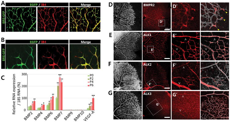
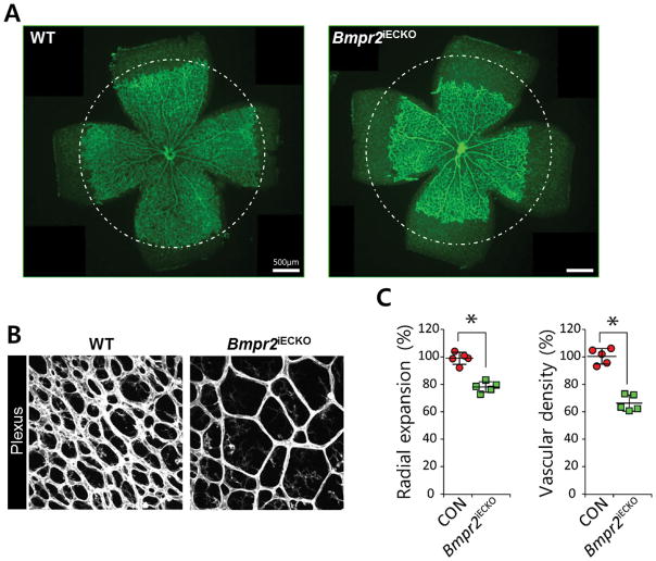
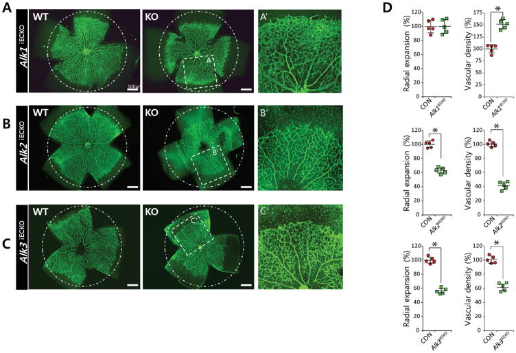
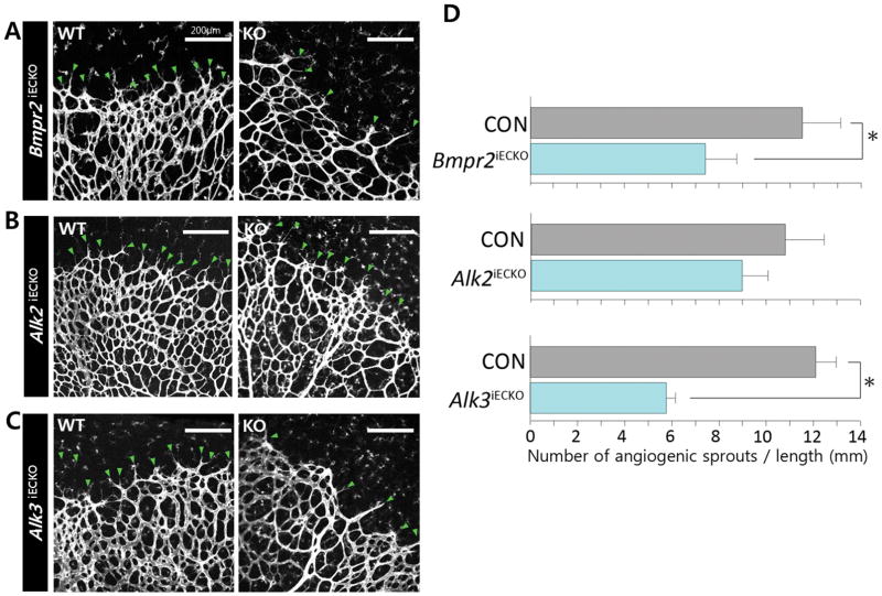
Approach and results: Expression analysis of several BMP ligands showed that proangiogenic BMP ligands are highly expressed in postnatal retinas. Consistently, BMP receptors are also strongly expressed in retina with a distinct pattern. To assess the function of BMP signaling in retinal angiogenesis, we first generated mice carrying an endothelial-specific inducible deletion of Bmpr2. Postnatal deletion of Bmpr2 in endothelial cells substantially decreased the number of angiogenic sprouts at the vascular front and branch points behind the front, leading to attenuated radial expansion. To identify critical BMPR1s (BMP type 1 receptors) associated with BMPR2 in retinal angiogenesis, we generated endothelial-specific inducible deletion of 3 BMPR1s abundantly expressed in endothelial cells and analyzed the respective phenotypes. Among these, endothelial-specific deletion of either Alk2/acvr1 or Alk3/Bmpr1a caused a delay in radial expansion, reminiscent of vascular defects associated with postnatal endothelial-specific deletion of BMPR2, suggesting that ALK2/ACVR1 and ALK3/BMPR1A are likely to be the critical BMPR1s necessary for proangiogenic BMP signaling in retinal vessels.

Conclusions: Our data identify BMP signaling mediated by coordination of ALK2/ACVR1, ALK3/BMPR1A, and BMPR2 as an essential proangiogenic cue for retinal vessels.

Keywords: BMP signaling; angiogenesis; retina; vertebrate development.


Figures

FIGURE 1
FIGURE 1. Diverse BMP ligands and receptors are expressed during retinal development
(A–B) Representative overview of retinal vessels at (A) the retinal center and (B) vascular front shown in higher magnification in postnatal day P5 BRE-gfp transgenic mice. Endothelial cells are shown in red. BRE-GFP is highly expressed in both tip cells and stalk cells. (C) qRT-PCR for Bmp ligands and Vegfa in P0, P2, P4, and P6 retina. Bmp6 and Bmp7 are the most abundant BMP ligands between P0 and P6 (n=5). (D–G) Immunohitochemistry of P5 retina showing BMPR2 (D), ALK1/ACVRL (E), ALK2/ACVR1 (F), and ALK3/BMPR1A (G). While BMPR2, ALK1 and ALK3 are expressed ubiquitously, ALK2 expression is enriched in the veins within the plexus region; scale bar, 300μm. (H–K) Areas within the white rectangles in panels D to G are shown in higher magnification.
FIGURE 2
FIGURE 2. BMPR2 is essential for retinal angiogenesis
(A) Representative overview of P5 retinal vessels taken from inducible endothelial specific knock out of Bmpr2fl/fl;Cdh5(Pac)CreERT2 (right) and their phenotypic wild-type littermates (left); scale bar, 500μm. Mean radial expansion of the retinal vessels in P5 Bmpr2fl/fl retinas was decreased to 78.9±9.8% of wildtype littermates. (B) Representative overview of plexus region of P5 retina in Bmpr2fl/fl;Cdh5(Pac)CreERT2 (right) and their phenotypic wild-type littermates (left). Mean vascular density in the plexus region in Bmpr2fl/fl retinas was reduced to 65.7±8.3% of wildtype littermates; scale bar, 500μm. Endothelial cells are visualized by anti- Isolectin B4 (IB4) staining. (C) Quantification of radial expansion and vascular density (% vascularized area) in Bmpr2fl/+;Cdh5(Pac)CreERT2 (gray) and Bmpr2fl/fl;Cdh5(Pac)CreERT2 mice injected with 100μg tamoxifen at P1, retinas were assayed P5 (n=5). P<0.05. Statistical significance was assessed using a Student’s unpaired t-test.
FIGURE 3
FIGURE 3. ALK2 and ALK3 promotes retinal angiogenesis
(AC) Representative overview of retinal vessels taken from inducible endothelial specific knock out of Alk1fl/fl;Cdh5(Pac)CreERT2 (A), Alk2fl/fl;Cdh5(Pac)CreERT2 (B), or Alk3fl/fl;Cdh5(Pac)CreERT2 (C) and their phenotypic wild-type littermates. Mice were injected with 50μg tamoxifen at P1, retinas were assayed P5; scale bar, 500μm. Both radial expansion and vascular density in the plexus region were significantly increased in P5 Alk1fl/fl;Cdh5(Pac)CreERT2 retinas (101±6.7% for radial expansion and 155±6.2% for vascular density compared to wildtype littermates). By contrast, deletion of either Alk2 or Alk3 significantly decreased both radial expansion (63.2±17.1% for Alk2fl/f;Cdh5(Pac)CreERT2 retinas and 56.4±14.2% for Alk3fl/fl;Cdh5(Pac)CreERT2 retinas compared to wildtype littermates) and vascular density in the plexus region (42.3±15.9% for Alk2fl/f;Cdh5(Pac)CreERT2 l retinas, and 61.9±12.8% for Alk3fl/fl;Cdh5(Pac)CreERT2 retinas compared to wildtype littermates). Areas within the white rectangle in middle column are shown in higher magnification (right column). While radial expansion was similarly affected by deletion of either Alk2 or Alk3, vascular density in the plexus region is more severely affected by the deletion of Alk2 than Alk3. Endothelial cells are visualized by anti- Isolectin B4 (IB4) staining. (D) Quantification of radial expansion and vascular density (% vascularized area) in inducible endothelial specific knockout of each BMPR1 (pink) and their littermates (gray) (n=5). P<0.05. Statistical significance was assessed using a Student’s unpaired t-test.
FIGURE 4
FIGURE 4. ALK3/BMPR2 signaling is required for the formation of angiogenic sprouts in the vascular front
(A) Representative image of vascular front in postnatal day P5 Bmpr2fl/fl;Cdh5(Pac)CreERT2 (A), Alk2fl/fl; Cdh5(Pac)CreERT2 (B), or Alk3fl/fl;Cdh5(Pac)CreERT2 Endothelial cells are visualized by anti- Isolectin B4 (IB4) staining. (C) and their phenotypic wild-type littermates. Green arrows point the angiogenic sprouts in the vascular front (n=5); scale bar, 200μm. (D) Quantification of angiogenic sprouts. The number of angiogenic sprouts in the vascular front was measured by dividing the number of angiogenic sprouts by the total length of vascular front (See Supplemental methods for detail). While the number of angiogenic sprouts in the vascular front was mildly decreased by 20% to in P5 Alk2fl/fl retinas (8.8±0.2 compared to 10.8±1.9 in wildtype littermates), it was decreased by 45% in Bmpr2fl/fl (6.5±1.3 in Bmpr2fl/fl retinas compared to 11.8±1.6 in wildtype littermates), and by 53% in Alk3fl/fl retinas (5.3±0.4 in Alk3fl/fl retinas compared to 11.2±0.9 in wildtype littermates). P<0.05. Statistical significance was assessed using a Student’s unpaired t-test.

References

    1. Cai J, Pardali E, Sanchez-Duffhues G, ten Dijke P. Bmp signaling in vascular diseases. FEBS letters. 2012;586:1993–2002. - PubMed
    1. David L, Feige JJ, Bailly S. Emerging role of bone morphogenetic proteins in angiogenesis. Cytokine & growth factor reviews. 2009;20:203–212. - PubMed
    1. Langenfeld EM, Langenfeld J. Bone morphogenetic protein-2 stimulates angiogenesis in developing tumors. Molecular cancer research : MCR. 2004;2:141–149. - PubMed
    1. Pi X, Ren R, Kelley R, Zhang C, Moser M, Bohil AB, Divito M, Cheney RE, Patterson C. Sequential roles for myosin-x in bmp6-dependent filopodial extension, migration, and activation of bmp receptors. The Journal of cell biology. 2007;179:1569–1582. - PMC - PubMed
    1. Wiley DM, Kim JD, Hao J, Hong CC, Bautch VL, Jin SW. Distinct signalling pathways regulate sprouting angiogenesis from the dorsal aorta and the axial vein. Nature cell biology. 2011;13:686–692. - PMC - PubMed

MeSH terms

Substances