. 2017 Apr 14;292(15):6339-6351.
doi: 10.1074/jbc.M116.746859. Epub 2017 Feb 23.

Ectodomain shedding of the cell adhesion molecule Nectin-4 in ovarian cancer is mediated by ADAM10 and ADAM17

Affiliations

Ectodomain shedding of the cell adhesion molecule Nectin-4 in ovarian cancer is mediated by ADAM10 and ADAM17

Petra C Buchanan et al. J Biol Chem. .

Abstract

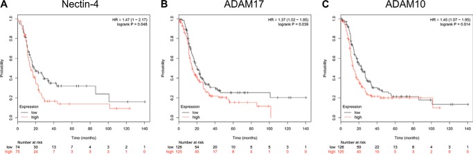
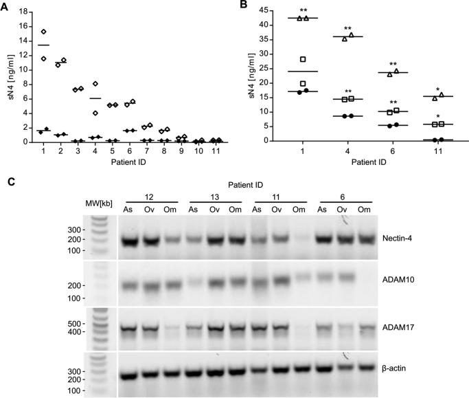
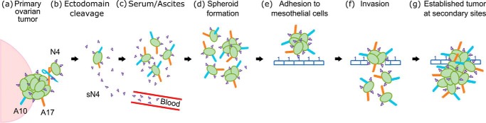
We previously showed that the cell adhesion molecule Nectin-4 is overexpressed in ovarian cancer tumors, and its cleaved extracellular domain can be detected in the serum of ovarian cancer patients. The ADAM (adisintegrin and metalloproteinase) proteases are involved in ectodomain cleavage of transmembrane proteins, and ADAM17 is known to cleave Nectin-4 in breast cancer. However, the mechanism of Nectin-4 cleavage in ovarian cancer has not yet been determined. Analysis of ovarian cancer gene microarray data showed that higher expression of Nectin-4, ADAM10, and ADAM17 is associated with significantly decreased progression-free survival. We quantified Nectin-4 shedding from the surface of ovarian cancer cells after stimulation with lysophosphatidic acid. We report that ADAM17 and ADAM10 cleave Nectin-4 and release soluble Nectin-4 (sN4). Small molecule inhibitors and siRNA knockdown of both ADAM proteases confirmed these results. In matched samples from 11 high-grade serous ovarian cancer patients, we detected 2-20-fold more sN4 in ascites fluid than serum. Co-incubation of ovarian cancer cells with ascites fluid significantly increased sN4 shedding, which could be blocked using a dual inhibitor of ADAM10 and ADAM17. Furthermore, we detected RNA for Nectin-4, ADAM10, and ADAM17 in primary ovarian carcinoma tumors, secondary omental metastases, and ascites cells isolated from serous ovarian cancer patients. In a signaling pathway screen, lysophosphatidic acid increased phosphorylation of AKT, EGF receptor, ERK1/2, JNK1/2/3, and c-Jun. Understanding the function of Nectin-4 shedding in ovarian cancer progression is critical to facilitate its development as both a serum biomarker and a therapeutic target for ovarian cancer.

Keywords: ADAM; ADAM10; ADAM17; Nectin-4; biomarker; cancer biology; cell migration; ovarian cancer.


Conflict of interest statement

The authors declare that they have no conflicts of interest with the contents of this article

Figures

Figure 1.
Figure 1.
Progression-free survival analysis of serous ovarian cancer patients. The Kaplan-Meier plotter for ovarian cancer was used to plot progression-free survival data of grade 1 and 2 serous ovarian cancer patients relative to gene expression. The program combines Affymetrix microarray data from the EGA and TCGA. The PFS data are shown for Nectin-4 (p = 0.048, A), ADAM17 (p = 0.039, B), and ADAM10 (p = 0.014, C). Lower expression is shown as a black line, and higher expression is shown as a red line. The respective Affymetrix IDs used for the calculations were 223540_at (Nectin-4), 213532_at (ADAM17), and 202604_x_at (ADAM10).
Figure 2.
Figure 2.
Flow cytometric analysis of NIH:OVCAR5 cells for expression of Nectin-4, ADAM17, and ADAM10. Confluent monolayers of cells were detached from tissue culture flasks with Accutase, washed, and labeled with mouse IgG or antibodies against Nectin-4, ADAM17, or ADAM10 (2.5 μg/106 cells) in flow buffer (PBS containing 2.5% newborn calf serum and 0.02% sodium azide). The cells were washed and incubated with goat anti-mouse IgG F(ab′)2, then washed again, and incubated with streptavidin-allophycocyanin (APC) conjugate. The cells were washed, fixed in flow buffer containing 1% formaldehyde, and analyzed by flow cytometry. NIH:OVCAR5 parental cells (A) and NIH:OVCAR5-Nectin-4 overexpressing cells (B) were analyzed for the expression of Nectin-4 (blue), ADAM17 (pink), ADAM10 (green), or mouse IgG negative control (open).
Figure 3.
Figure 3.
The role of ADAM17 and ADAM10 in Nectin-4 ectodomain shedding. A and B, NIH:OVCAR5-N4-over cells were seeded in 24-well plates and after 24 h in culture, they were stimulated with 100 ng/ml PMA (A, white triangles) or 10 μm LPA (B, white squares) in OptiMem for 1, 3, or 6 h at 37 °C. Constitutive shedding without stimulation was also analyzed (black circles). Supernatants were collected, and shed N4 (sN4) was quantified in triplicate by ELISA. Shown is a representative of three independent experiments. Error bars, S.D. C and D, in another assay, NIH:OVCAR5-N4-over cells were seeded in 24-well plates and incubated with the respective broad-spectrum or selective ADAM sheddase inhibitor (Table 1) for 30 min before stimulating with 100 ng/ml PMA (C, white triangles) or 10 μm LPA (D, white triangles) for 3 h at 37 °C. Inhibition of constitutive shedding without stimulation was also analyzed (black circles). Supernatants were collected, and shed N4 was quantified in triplicate by ELISA. Shown is a representative of three independent experiments. The mean of the replicates is shown as a solid line; the unpaired Student's t test was used to calculate significant inhibition of shedding by the inhibitors used (**, p < 0.01; *, p < 0.05); each sample with inhibitor was compared against the respective untreated control sample (white triangles, W/O).
Figure 4.
Figure 4.
siRNA-mediated knockdown of ADAM proteases. NIH:OVCAR5-N4-over cells were seeded in 24-well plates and after 24 h in culture, they were transfected with 25 nm of Dharmacon On-Targetplus siRNA oligonucleotides: an siRNA negative control pool, an siRNA GAPDH control pool, an siRNA ADAM17 pool, an siRNA ADAM10 pool, or an siRNA pool targeting both ADAM proteases. In addition, an untreated control without siRNA transfection was prepared. Total cellular RNA was extracted 48 h after transfection, and 50 ng of RNA was analyzed by duplex RT-PCR (for GAPDH plus ADAM10 or ADAM17). Amplification products were visualized on a 0.9% agarose gel. The duplex RT-PCR for GAPDH plus ADAM17 (A) and GAPDH plus ADAM10 (B). Amplimer sizes: GAPDH (452 bp), ADAM10 (223 bp), and ADAM17 (304 bp) (see primers in Table 2). siRNA-treated cells (K.D.) were incubated with the inhibitors GI254023X (GI), BMS566395 (BMS), or INCB3619 (INCB) (Table 1) for 30 min before stimulating with: 100 ng/ml PMA (black triangles, C) or 10 μm LPA (white triangles, D) for 3 h at 37 °C. The supernatants were collected, and shed N4 was quantified by ELISA in duplicate. Shedding was quantified as a percentage of the shedding observed with the negative control siRNA pool. Shown is a representative of two independent experiments. The mean of the replicates is shown as a solid line. Student's t test unpaired shows significant inhibition of shedding (**, p < 0.01; *, p < 0.05); each knockdown sample was compared against the GAPDH control and the knockdown sample plus inhibitor (asterisks over brackets).
Figure 5.
Figure 5.
Quantification of shed Nectin-4 in patient biospecimens. A, matched serum (black diamonds) and ascites samples (white diamonds) of 11 high-grade serous ovarian cancer patients were analyzed for the amount of Nectin-4 by ELISA, in duplicate. Shown is a representative of two independent experiments. B, quantification of Nectin-4 shedding by cells incubated in patients' ascites fluid. NIH:OVCAR5-N4-over cells were seeded in 24-well plates and stimulated with ascites fluid (diluted 1:10 in OptiMem) from four high-grade serous ovarian cancer patients for 3 h at 37 °C (white triangles) or incubated with the ADAM10/ADAM17 inhibitor INCB3619 for 30 min before stimulating with patients' ascites fluid (white squares). Ascites fluid without cells (black circles) was also tested as a control for endogenous levels of sN4. Supernatants were collected, and sN4 was quantified by ELISA. Shown is a representative of two independent experiments. The unpaired Student's t test was used to calculate significant stimulation of shedding by incubation in ascites fluid (white triangles) or significant inhibition of shedding with INCB3619 (white squares) (**, p < 0.01; *, p < 0.05). C, RT-PCR of matched patient biospecimens. Ascites cells (As), primary ovarian tumor tissue (Ov), and omentum tissue (Om) of four high-grade serous ovarian cancer patients were analyzed for expression of Nectin-4, ADAM10, and ADAM17 by RT-PCR. The picture shows the 1% agarose gel electrophoresis of the samples with β-actin as a loading control. Lanes irrelevant to this study were removed from the figure.
Figure 6.
Figure 6.
LPA stimulated phosphorylation of signaling molecules in NIH:OVCAR5 cells with and without Nectin-4 expression. NIH:OVCAR5 cells with endogenous Nectin-4 expression (control, Ctrl) or NIH:OVCAR5 shRNA-mediated Nectin-4 knockdown cells (N4-KD) were analyzed using the Proteome Profiler human phospho-RTK and phosphokinase antibody arrays (circles, control cells; triangles, N4-KD cells). The cells were serum-starved overnight and either left untreated (filled symbols) or treated with 10 μm LPA for 1 h (open symbols) prior to preparation of protein extracts and incubation with antibody arrays. The density (in pixels) of the array spots is plotted for Akt1/2/3 (Ser-473, A); Akt1/2/3 (Thr-308, B); EGFR (phosphotyrosine, C); ERK1/2 (Thr-202/Tyr-204, Thr-185/Tyr-187, D); JNK1/2/3 (Thr-183/Tyr-185, Thr-221/Tyr-223, E); and c-Jun (Ser-63, F).
Figure 7.
Figure 7.
Model for ovarian cancer progression in which Nectin-4 plays a critical role. a, ovarian cancer cells (green solid ovals) express Nectin-4 (purple fours), ADAM10 (blue rod), and ADAM17 (orange rod) on their surface (Refs. , , and and this study). b, the ectodomain of Nectin-4 can be cleaved from the surface of ovarian cancer cells by ADAM10 and ADAM17 (this study). c, shed Nectin-4 can be detected in ascites fluid of ovarian cancer patients (this study) and in the serum of ovarian cancer patients (Ref. and this study). d, Nectin-4 expressing ascites cells can be found in patients (this study) and Nectin-4 is essential for the formation of compact spheroids (15). e, ovarian cancer spheroids adhere to Nectin-1 (blue ones) on mesothelial cells (blue rectangles) (4, 15). f, ovarian cancer spheroids can then disaggregate and invade mesothelial cell layers (5, 6). g, the presence of Nectin-4, ADAM10, and ADAM17 can be detected at secondary tumor sites (omental tissue in this study). Our data from this study and previous work implies that ovarian cancer cells strongly rely upon Nectin-4-mediated cell-cell interactions within the peritoneal cavity to metastasize.

Similar articles

Cited by

References

    1. Siegel R. L., Miller K. D., and Jemal A. (2015) Cancer statistics, 2015. CA Cancer J. Clin. 65, 5–29 - PubMed
    1. Perets R., and Drapkin R. (2016) It's totally tubular: riding the new wave of ovarian cancer research. Cancer Res. 76, 10–17 - PMC - PubMed
    1. Pradeep S., Kim S. W., Wu S. Y., Nishimura M., Chaluvally-Raghavan P., Miyake T., Pecot C. V., Kim S. J., Choi H. J., Bischoff F. Z., Mayer J. A., Huang L., Nick A. M., Hall C. S., Rodriguez-Aguayo C., et al. (2014) Hematogenous metastasis of ovarian cancer: rethinking mode of spread. Cancer Cell 26, 77–91 - PMC - PubMed
    1. Burleson K. M., Casey R. C., Skubitz K. M., Pambuccian S. E., Oegema T. R. Jr., and Skubitz A. P. (2004) Ovarian carcinoma ascites spheroids adhere to extracellular matrix components and mesothelial cell monolayers. Gynecol. Oncol. 93, 170–181 - PubMed
    1. Burleson K. M., Hansen L. K., and Skubitz A. P. (2004) Ovarian carcinoma spheroids disaggregate on type I collagen and invade live human mesothelial cell monolayers. Clin. Exp. Metastasis 21, 685–697 - PubMed

MeSH terms