. 2017 Feb 24;355(6327):eaag1789.
doi: 10.1126/science.aag1789.

The [4Fe4S] cluster of human DNA primase functions as a redox switch using DNA charge transport

Affiliations

The [4Fe4S] cluster of human DNA primase functions as a redox switch using DNA charge transport

Elizabeth O'Brien et al. Science. .

Abstract

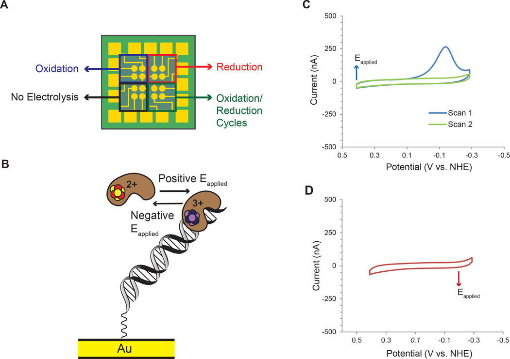
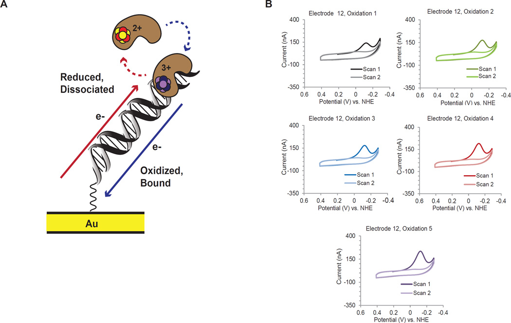
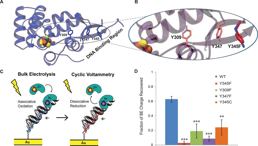
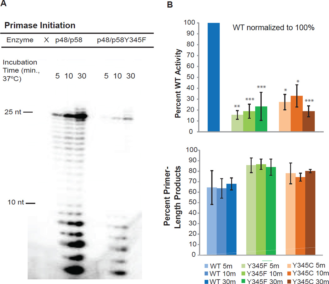
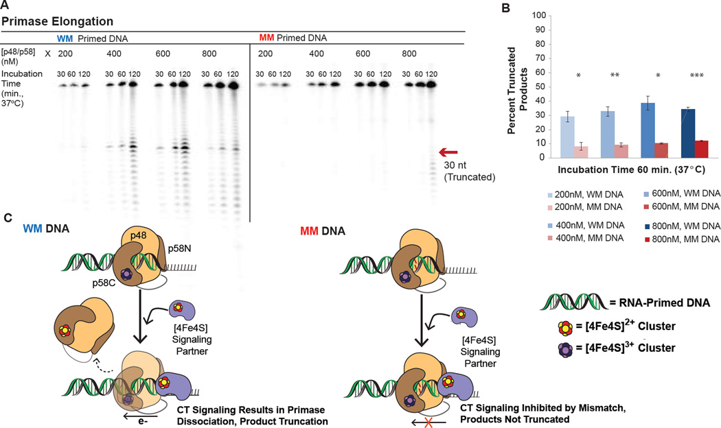
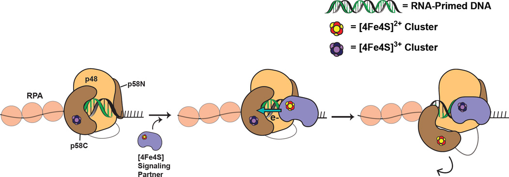
DNA charge transport chemistry offers a means of long-range, rapid redox signaling. We demonstrate that the [4Fe4S] cluster in human DNA primase can make use of this chemistry to coordinate the first steps of DNA synthesis. Using DNA electrochemistry, we found that a change in oxidation state of the [4Fe4S] cluster acts as a switch for DNA binding. Single-atom mutations that inhibit this charge transfer hinder primase initiation without affecting primase structure or polymerization. Generating a single base mismatch in the growing primer duplex, which attenuates DNA charge transport, inhibits primer truncation. Thus, redox signaling by [4Fe4S] clusters using DNA charge transport regulates primase binding to DNA and illustrates chemistry that may efficiently drive substrate handoff between polymerases during DNA replication.


Figures

Figure 1
Figure 1. Oxidized [4Fe4S]3+ and reduced [4Fe4S]2+ p58C display different behavior on DNA-modified electrodes
A) Multiplex chip with 16 DNA-modified Au electrodes (circles, center.) This platform facilitates direct comparison of oxidized, reduced, unaltered, and iteratively oxidized samples, on four separate quadrants of a single surface. B) The cartoon depicts the effects of electrochemical oxidation and reduction on p58C DNA binding and redox activity. C) CV of electrochemically oxidized p58C. After electrochemical conversion (Eapplied = 412mV vs. NHE) of the sample at the electrode/solution interface to the [4Fe4S]3+ state, CV scans display a large cathodic peak only in the initial scan to negative, reducing potentials. D) CV of electrochemically reduced p58C. After electrochemical conversion (Eapplied = −188 mV vs. NHE) of the sample to the [4Fe4S]2+ state, CV scans show no electrochemical signal on DNA. Electrochemistry was performed on 16µM p58C in 20 mM Tris, pH 7.2, 75 mM NaCl, 100mV/s for CV scans, using a Ag/AgCl reference electrode.
Figure 2
Figure 2. Iterative oxidation/reduction cycles of p58C on a single electrode surface
A) The scheme depicts the redox switch in p58C DNA binding. When oxidized, p58C is bound tightly to DNA. Reduction converts p58C to a weakly DNA-associated state. B) Cyclic voltammetry following five sequential oxidation reactions on one DNA-modified electrode of a multiplex chip. Electrolysis conditions (Eapplied = 412mV vs. NHE) are identical for each oxidation. A cathodic peak at 130- 140mV vs. NHE is regenerated each time in the first CV scan after oxidation. The cathodic peak corresponds to a reduction of tightly bound, oxidized p58C, to weakly associated p58C. Charge transfer values in the cathodic peaks for scans 1–5, in chronological order, are 65.4 nC, 112.4 nC, 116.4 nC, 151.1 nC, and 170.9 nC. Peak charge increases over trials due to increasing p58C at the solution/DNA interface. Electrochemistry was performed on 16 µM p58C in 20 mM Tris, pH 7.2, 75 mM NaCl, 100 mV/s scan rate for CV, using a Ag/AgCl reference electrode.
Figure 3
Figure 3. DNA-binding, charge-transfer deficient p58C mutants
A) Tyrosine residues conserved in eukaryotic primase [4Fe4S] domain (Y309, Y345, Y347 in H. sapiens, blue sticks) are located between the [4Fe4S] cluster (orange and yellow spheres) and DNA-binding region. The DNA binding region, consisting of residues R302, R306, K314, and W327, is located ~20–30 Å from the cluster, necessitating electron transfer through the protein matrix for exchange of charge between the [4Fe4S] cofactor and bound DNA. B) Expanded region of the overlaid crystal structures of p58C (PDB 3L9Q, blue) and p58C Y345F (PDB 517M, red) demonstrates the minimal structural impact of the Y-F mutation; the phenylalanine residue in the mutant adapts the same orientation as the tyrosine in WT p58C. All mutants bind DNA with approximately the same affinity as WT p58C. C) Scheme depicts redox reactions in electrochemical assays with wild type and mutant p58C. Bulk electrolysis first oxidizes p58C and promotes tight DNA binding. CV then reduces the DNA-bound protein, converting it to the weakly associated, electrochemically inactive form. Both require the tyrosine charge transfer pathway and must be accounted for when comparing charge transfer proficiency. D) WT p58C recovers significantly more (63±4%) bulk electrolysis charge than the mutants, suggesting that perturbation of the charge transfer pathway diminishes DNA-bound redox chemistry and consequently affects the redox switch. All bulk electrolysis reactions and CV scans were performed on 16 µM p58C/mutant in 20 mM Tris, pH 7.2, 75 mM NaCl at a 100 mV/s scan rate for CV, using a Ag/AgCl reference electrode. Mean ± SD of n=3 scans per variant, ** = 0.001<p<0.0005, *** = p<0.0005, student’s t-test.
Figure 4
Figure 4. Redox Switching Plays a Role in Primase Initiation
A) Gel separation of products for WT p48/p58 and p48/p58Y345F reactions on ssDNA. The WT enzyme is significantly more active on ssDNA than either mutant. B) Quantified products for WT p48/p58, p48/p58Y345F, and p48/p58Y345C initiation assays. Mutants synthesize 15–35% of WT products on average. Mutant primase synthesizes shorter products on average. Primer-length products in graph below are defined as products 7–10nt in length. Initiation assays were performed anaerobically, with 250nM ssDNA, 1 µM α-32P ATP, 112 µM CTP, 188 µM UTP, 400 nM enzyme in 50 mM Tris, pH 8.0, 3 mM MgCl2, at 37 °C. Quantifications shown are mean ± SD of n ≥ 3 trials, * = 0.001<p<0.005, ** = 0.001<p<0.0005, *** = p<0.0005, student’s t-test.
Figure 5
Figure 5. A Mismatch in the Nascent Primer Inhibits Primase Truncation
A) Gel separation of elongation products on a 2’-OMe RNA- primed DNA substrate, when a well-matched (WM) or mismatched (MM) primer is synthesized by WT p48/p58. B) Average percent truncated products after 60 minutes of incubation at 37°C. WT primase synthesizes significantly more truncated products with a WM primer than a MM primer. C) Scheme illustrating the observed products in the mismatched primer elongation experiment. When p58C is in contact with the RNA/DNA primer, primase can signal another DNA-bound [4Fe4S] enzyme through a WM primer and dissociate from the template, truncating products. bottom left) DNA CT is inhibited with a MM primer, precluding redox signaling and primer truncation. Elongation assays were performed anaerobically, with 500nM primed DNA, 1 µM α-32P ATP, 200 µM CTP, 100 µM UTP, 200–800 nM p48/p58 in 50 mM Tris, pH 8.0, 3 mM MgCl2, at 37 °C. Quantifications shown are mean ± SD of n = 3 trials, * = 0.001<p<0.005, ** = 0.001<p<0.0005, *** = p<0.0005, student’s t-test.
Figure 6
Figure 6. Proposed Mechanism of Primer Handoff Driven by DNA Charge Transport Chemistry
DNA primase elongates an RNA primer (green) to 8–12 nt length with both p48 and p58C contacting the nascent RNA/DNA duplex (left). When the nascent primer is large enough, another [4Fe4S] enzyme (purple), which we hypothesize to be DNA polymerase αin vivo, participates in DNA-mediated signaling through the primer-template duplex (middle). This promotes dissociation of p58C through reduction of the cluster from the [4Fe4S]3+ state to the [4Fe4S]2+ state; the next [4Fe4S] enzyme is then tightly bound and can continue elongation of the primer-template (right).

Comment in

References

    1. Grodick MA, Muren NB, Barton JK. DNA charge transport within the cell. Biochemistry. 2015;54:962–973. - PMC - PubMed
    1. Gorodetsky AA, Boal AK, Barton JK. Direct electrochemistry of endonuclease III in the presence and absence of DNA. J. Am. Chem. Soc. 2006;128:12082–12083. - PubMed
    1. Boal AK, Genereux JC, Sontz PA, Gralnick JA, Newman DK, Barton JK. Redox signaling between DNA repair proteins for efficient lesion detection. Proc. Natl. Acad. Sci. USA. 2009;106:15237–15242. - PMC - PubMed
    1. Sontz PA, Mui TP, Fuss JO, Tainer JA, Barton JK. DNA charge transport as a first step in coordinating the detection of lesions by repair proteins. Proc. Natl. Acad. Sci. USA. 2012;109:1856–1861. - PMC - PubMed
    1. Pheeney CG, Arnold AR, Grodick MA, Barton JK. Multiplexed electrochemistry of DNA-bound metalloproteins. J. Am. Chem. Soc. 2013;135:11869–11878. - PMC - PubMed

Publication types