. 2017 Feb 24;49(2):e296.
doi: 10.1038/emm.2016.151.

Resveratrol suppresses breast cancer cell invasion by inactivating a RhoA/YAP signaling axis

Affiliations

Resveratrol suppresses breast cancer cell invasion by inactivating a RhoA/YAP signaling axis

Yu Na Kim et al. Exp Mol Med. .

Abstract

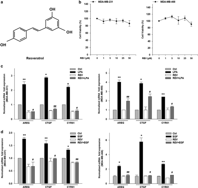
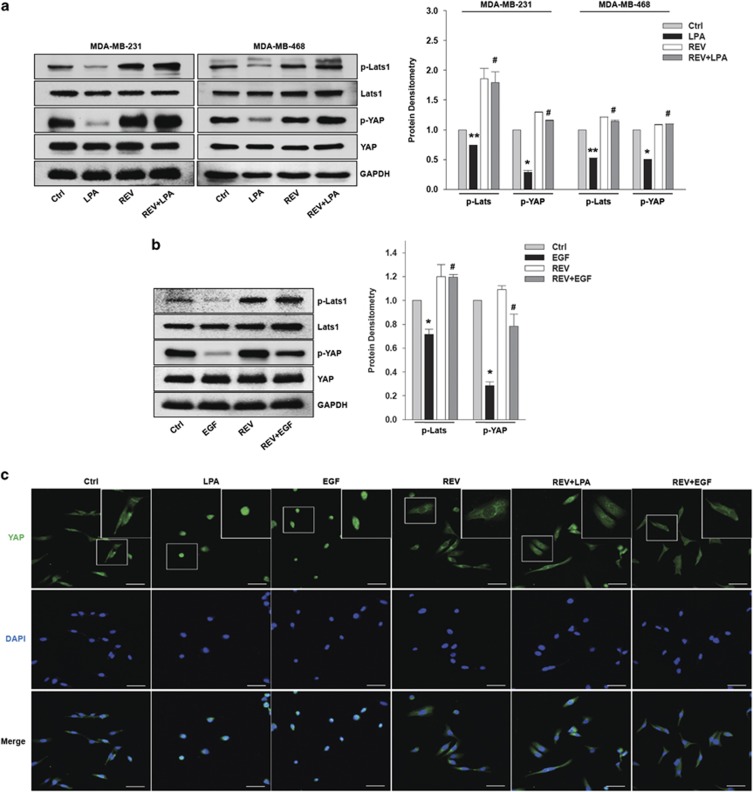
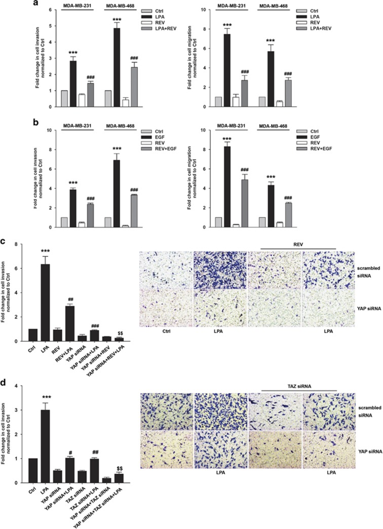
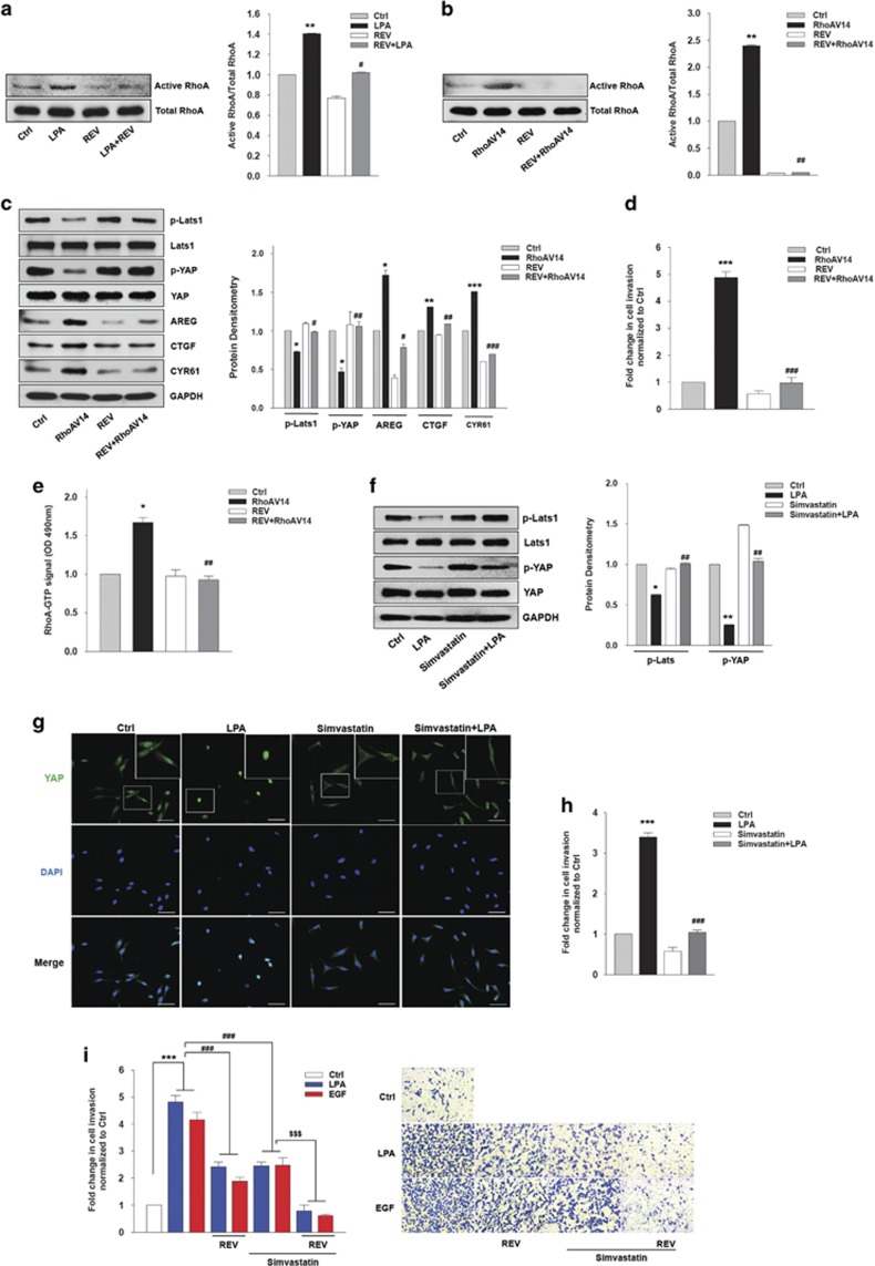
Hippo/YAP signaling is implicated in tumorigenesis and progression of various cancers. By inhibiting a plethora signaling cascades, resveratrol has strong anti-tumorigenic and anti-metastatic activity. In the present study, we demonstrate that resveratrol decreases the expression of YAP target genes. In addition, our data showed that resveratrol attenuates breast cancer cell invasion through the activation of Lats1 and consequent inactivation of YAP. Strikingly, we also demonstrate that resveratrol inactivates RhoA, leading to the activation of Lats1 and induction of YAP phosphorylation. Further, resveratrol in combination with other agents that inactivate RhoA or YAP showed more marked suppression of breast cancer cell invasion compared with single treatment. Collectively, these findings indicate the beneficial effects of resveratrol on breast cancer patients by suppressing the RhoA/Lats1/YAP signaling axis and subsequently inhibiting breast cancer cell invasion.


Conflict of interest statement

The authors declare no conflict of interest.

Figures

Figure 1
Figure 1
Resveratrol (REV) reduces the expression of yes-associated protein target genes. (a) The chemical structure of REV. (b) The viability of breast cancer cell was analyzed by (3,4,5-dimethylthiazol-2yl)-5-diphenyl-tetrazolium bromide (MTT) assays. The serum-starved cells were treated with REV for 24 h. (c, d) The serum-starved MDA-MB-231 and MDA-MB-468 cells were pretreated with REV (25 μM) for 1 h and then stimulated with lysophosphatidic acid (LPA; 5 μM) and epidermal growth factor (EGF; 100 ng ml−1) for 2 and 0.5 h, respectively. Quantitative reverse transcription-PCR (*P<0.05 and **P<0.01 versus control, #P<0.05 and ##P<0.01 versus LPA or epidermal growth factor (EGF) treatment only). All experiments were repeated three times.
Figure 2
Figure 2
Resveratrol (REV) inhibits lysophosphatidic acid (LPA)- and epidermal growth factor (EGF)-induced YAP activation. (a–c) The serum-starved MDA-MB-231 and MDA-MB-468 cells were pretreated with REV (25 μM) for 1 h and then stimulated with LPA (5 μM) and EGF (100 ng ml−1) for 2 h and 0.5 h, respectively. (a, b) Immunoblotting. Immunoblot bands were quantified by ImageJ densitometric analysis and normalized to Lats1 and YAP, respectively (*P<0.05, **P<0.01 versus control, #P<0.05 versus LPA and EGF treatment only). (c) Immunofluorescence. Original magnification × 200; scale bar=50 μm. All experiments were repeated three times.
Figure 3
Figure 3
Resveratrol (REV) suppresses lysophosphatidic acid (LPA)- and epidermal growth factor (EGF)-induced breast cancer cell invasion. (a, b) The serum-starved MDA-MB-231 and MDA-MB-468 cells were pretreated with REV (25 μM) for 1 h and in vitro invasion and migration were analyzed against LPA (5 μM) (a) and EGF (100 ng ml−1) (b) (***P<0.001 versus control, ###P<0.001 versus LPA and EGF treatment only). (c) The MDA-MB-231 cells were transfected with the indicated siRNA. Then, the cells were pretreated with REV (25 μM) for 1 h, followed by stimulation with or without LPA (5 μM; ***P<0.001 versus control, ##P<0.01 versus LPA treatment only, ###P<0.001 versus LPA treatment only, $$P<0.01 versus LPA treatment and YAP siRNA). Original magnification × 200. (d) The MDA-MB-231 cells were transfected with the indicated siRNA and then stimulated with or without LPA (5 μM; ***P<0.001 versus control, #P<0.05 versus LPA treatment only, ##P<0.01 versus LPA treatment only, $$P<0.01 versus TAZ siRNA and LPA treatment). Original magnification of all images, × 200. All experiments were repeated three times.
Figure 4
Figure 4
Resveratrol (REV) inactivates RhoA. (a) The serum-starved MDA-MB-231 cells were pretreated with REV (25 μM) and then stimulated with lysophosphatidic acid (LPA; 5 μM) for 3 min. Active RhoA bands were quantified by ImageJ densitometric analysis and normalized to total RhoA (**P<0.01 versus control, #P<0.05 versus LPA treatment). (b–e) The MDA-MB-231 cells were transfected with the indicated vectors for 48 h and then treated with REV (25 μM) for 1 h. (b) RhoA-GTP pull-down assays. Active RhoA bands were quantified by ImageJ densitometric analysis and normalized to total RhoA (**P<0.01 versus control, ##P<0.05 versus transfection of RhoAV14). (c) Immunoblotting. Immunoblot bands were quantified by ImageJ densitometric analysis and normalized to the control (*P<0.05 versus control vector, **P<0.01 versus control vector, ***P<0.001 versus control vector, #P<0.05 versus transfection of RhoAV14, ##P<0.01 versus transfection of RhoAV14, ###P<0.001 versus transfection of RhoAV14). (d) In vitro invasion assay (***P<0.001 versus control vector, ###P<0.001 versus transfection of RhoAV14). (e) RhoA GLISA activation assay (*P<0.05 versus control, ##P<0.01 versus V14 RhoA treatment only). (f–h) The serum-starved MDA-MB-231 cells were pretreated with simvastatin (5 μM) for 1 h and then stimulated with LPA (5 μM) for 2 h. (f) Immunoblotting. Immunoblot bands were quantified by ImageJ densitometric analysis and normalized to Lats1 and YAP (*P<0.05, **P<0.01 versus control, ##P<0.01 versus LPA treatment only). (g) The expression of YAP was visualized by immunofluorescence. Original magnification × 200; scale bar, 50 μm. (h) In vitro invasion assay (***P<0.001 versus control, ###P<0.001 versus LPA treatment only). (i) The serum-starved MDA-MB-231 cells were pretreated with REV (25 μM) or simvastatin (5 μM) for 1 h and in vitro invasion was analyzed against LPA (5 μM) or epidermal growth factor (EGF; 100 ng ml−1; ***P<0.001 versus control, ###P<0.001 versus LPA or EGF treatment only, $$$P<0.001 versus LPA or EGF and simvastatin treatment). All experiments were repeated three times.

References

    1. Pan D. The hippo signaling pathway in development and cancer. Dev Cell 2010; 19: 491–505. - PMC - PubMed
    1. Harvey KF, Zhang X, Thomas DM. The Hippo pathway and human cancer. Nat Rev Cancer 2013; 13: 246–257. - PubMed
    1. Zhao B, Tumaneng K, Guan KL. The Hippo pathway in organ size control, tissue regeneration and stem cell self-renewal. Nat Cell Biol 2011; 13: 877–883. - PMC - PubMed
    1. Kilili GK, Kyriakis JM. Mammalian Ste20-like kinase (Mst2) indirectly supports Raf-1/ERK pathway activity via maintenance of protein phosphatase-2A catalytic subunit levels and consequent suppression of inhibitory Raf-1 phosphorylation. J Biol Chem 2010; 285: 15076–15087. - PMC - PubMed
    1. Praskova M, Xia F, Avruch J. MOBKL1A/MOBKL1B phosphorylation by MST1 and MST2 inhibits cell proliferation. Curr Biol 2008; 18: 311–321. - PMC - PubMed

MeSH terms